
Early Brain Health Care Can Save Lives: 12 Warning Signs
Early Brain Health Care Can Save Lives: 12 Warning Signs You Should Never Ignore Brain Health Care is not something most people think about until a symptom forces them to. Yet the truth is simple: the earlier a neurological problem is caught, the better the outcome tends to be. Many serious neurological disorders — from stroke to Alzheimer’s disease — become far more difficult to treat once diagnosis is delayed, and in some cases, that delay can mean the difference between a full recovery and permanent disability. At Consultant Corner, we see this pattern again and again. A patient waits a few days to “see if the numbness goes away.” A family assumes memory slips are “just aging.” A headache is dismissed as stress. In many cases, nothing serious is happening. But in the cases where something is wrong, that waiting period is exactly when the most damage occurs. This is why proactive brain health care — built around prompt evaluation and fast scheduling — is one of the most important things you can do for your long-term independence and quality of life. In this guide, we’ll walk through the 12 warning signs that call for immediate neurological evaluation, explain why timing matters so much in neurology, and show you how Consultant Corner supports patients across Illinois and Texas with rapid access to expert care. Why Brain Health Care Depends on Speed Unlike many areas of medicine, neurology is often a race against the clock. Brain tissue is extremely sensitive to interruptions in blood flow and oxygen, and nerve damage can become permanent within a matter of hours. This is why brain health care providers emphasize early evaluation over a “wait and see” approach. Consider ischemic stroke, one of the most time-sensitive emergencies in all of medicine. When a blood vessel supplying the brain becomes blocked, brain cells begin to die within minutes. Our detailed guide on Ischemic Stroke: Early Warning Signs, Treatment and Recovery explains how quickly treatment decisions need to be made and why every minute of delay can cost brain function that may never fully return. The same principle applies — just on a different timeline — to conditions like Alzheimer’s disease, chronic migraines, epilepsy, and nerve pain. The disease process may move slower, but early brain health care still changes the trajectory. Catching a problem in its earliest stage typically means: Fewer complications A wider range of treatment options Better response to medication and therapy Slower disease progression Greater independence over the long term The Cost of Delayed Diagnosis When neurological symptoms are ignored or misattributed to stress, aging, or “being tired,” patients often lose the window where treatment is most effective. A tremor dismissed for years as nervousness may turn out to be an early, very treatable form of essential tremor — something we cover in depth in Essential Tremor Treatment: 10 Expert Tips to Stop the Shaking. A “few dizzy spells” may be an inner-ear issue — or it may be something more serious, as explained in Vertigo & Dizziness: 9 Common Causes. The pattern is consistent: symptoms that seem minor in isolation can be early signals of a much larger issue. That is precisely why brain health care should never be reactive-only. It should be proactive. 12 Warning Signs That Call for Immediate Neurological Evaluation Below are the 12 symptoms our neurology team most frequently identifies as red flags. If you or a loved one experiences any of these, it’s time to prioritize brain health care and schedule an evaluation right away. 1. Sudden Weakness Sudden weakness, especially on one side of the body — a drooping face, a weak arm, or a leg that suddenly gives out — is one of the classic warning signs of stroke. It should always be treated as an emergency. 2. Difficulty Speaking Slurred speech, trouble finding words, or sudden confusion when trying to communicate can signal a stroke, TIA (mini-stroke), or another acute neurological event. Our article on TIA (Mini-Stroke) Warning Signs You Should Learn explains why these episodes are easy to miss — and why they demand urgent follow-up even if symptoms resolve on their own. 3. Vision Loss Sudden blurred vision, double vision, or partial vision loss in one or both eyes can point to a vascular event in the brain or an underlying neurological condition that requires prompt imaging and evaluation. 4. Severe Headaches A headache that is sudden, unusually intense (“the worst headache of your life”), or paired with other neurological symptoms is different from an everyday tension headache. Our guide, Understanding Headache / Migraine, breaks down which headache patterns are ordinary and which ones warrant a same-week appointment. 5. Confusion Sudden confusion, disorientation, or an inability to follow a conversation can be a sign of stroke, seizure activity, or a metabolic or neurological emergency. This symptom is frequently underestimated by families who assume it’s temporary. 6. Loss of Balance Trouble walking, a sudden loss of coordination, or a feeling that the room is spinning can stem from several different neurological causes. If it happens suddenly and doesn’t resolve quickly, it deserves the same urgency as other stroke symptoms. 7. Seizures A first-time seizure — even a brief one — should always prompt a full neurological workup. Recurring seizures are a hallmark of epilepsy, and early diagnosis dramatically improves how well the condition can be managed long term. 8. Memory Decline Occasional forgetfulness is normal. But memory decline that interferes with daily routines, familiar tasks, or conversations may be an early sign of Alzheimer’s disease. Our related resource, Understanding Alzheimer’s Disease, outlines the earliest changes families should watch for. 9. Frequent Falls Falling more often than usual, especially without an obvious cause, can point to problems with balance, muscle strength, or nerve signaling. Our post on 8 Muscle Weakness Signs That May Need ALS Screening explains how progressive weakness and falls can sometimes be connected to a more serious underlying condition. 10. Persistent Numbness Numbness or tingling that doesn’t go away
Brain Health Care in Texas: 10 Warning Signs
Brain Health Care in Texas: 10 Warning Signs You Shouldn’t Ignore Brain health care is something most Texans don’t think about until a symptom forces the issue — a sudden headache that won’t quit, a hand that won’t stop shaking, or a parent who suddenly can’t remember the way home. But it isn’t just for emergencies. It’s an ongoing part of staying well, and across Texas, millions of people need this kind of neurological attention every single year. As the state’s population grows and ages, neurological conditions are becoming one of the most common reasons people see a specialist. Headaches, stroke, memory loss, Parkinson’s disease, neuropathy, seizures, dizziness, and spinal nerve disorders affect people in every corner of the state, from major metro areas to small rural towns. So many of these conditions develop slowly and quietly, which is exactly why a proactive approach built around early detection can be the difference between a manageable diagnosis and a life-altering one. This guide walks through why this matters so much right now, the ten warning signs every Texan should know, a closer look at the most common neurological conditions behind them, practical guidance on when a symptom is an emergency versus something to schedule, and how to find the right specialist — no matter where in Texas you live. Along the way, we’ll also cover the diagnostic tools neurologists rely on and answer some of the questions patients ask most often. Why Brain Health Care Matters More Than Ever in Texas Texas is growing fast, and its population is aging right along with it. That combination — more people, living longer — means more neurological care needs across the board every single year. Neurological disease doesn’t discriminate by zip code, but it does become more common with age, and Texas now has one of the largest populations of older adults in the country. Rural counties face an added challenge: fewer specialists per capita, which often means longer travel times and longer waits before someone with a developing condition ever sees a neurologist. The numbers help explain why. Nationally, someone in the United States has a stroke roughly every 40 seconds, and more than 795,000 people experience one every year. Texas ranks third in the nation for the number of residents living with Alzheimer’s disease, according to the Alzheimer’s Association’s Facts and Figures report, with hundreds of thousands of Texans over 65 currently affected. Migraine, epilepsy, Parkinson’s disease, and peripheral neuropathy add millions more to that total when you count every Texan living with a chronic neurological condition. None of this means every headache is a stroke or every forgotten name is Alzheimer’s. But it does mean that regular checkups, early screening, and fast attention to new symptoms deserve the same priority Texans already give to heart health or cancer screening — and that brain health care belongs in that same conversation. 10 Warning Signs That Call for Brain Health Care Many neurological diseases don’t announce themselves with a single dramatic event. Instead, they build gradually, which is exactly why early diagnosis is so important. The table below breaks down ten common warning signs, what they can indicate, and how urgently they should be evaluated. Warning Sign What It May Indicate How Urgent? Sudden weakness (face, arm, or leg) Stroke, TIA (mini-stroke) Emergency — call 911 Persistent headaches Migraine, tension headache, or secondary causes See a doctor within days Memory loss Alzheimer’s disease, other dementia Schedule an evaluation Tremors Essential tremor, Parkinson’s disease Schedule an evaluation Numbness or tingling Neuropathy, stroke, spinal nerve compression Same-day to same-week Balance problems Vertigo, cerebellar or inner-ear disorders Schedule an evaluation Speech difficulty Stroke, TIA Emergency — call 911 Vision changes Migraine, stroke, optic nerve issues Same-day evaluation Chronic back or neck pain Spinal nerve disorders, disc disease Schedule an evaluation Muscle weakness ALS, neuropathy, muscular disorders Schedule an evaluation If a symptom appears suddenly — especially sudden weakness, speech trouble, or vision loss — treat it as an emergency. For everything else on this list, a proactive approach to your neurological health works best when it starts early, before a gradual issue becomes a permanent one. A Closer Look at the Conditions Behind These Symptoms Ischemic Stroke Ischemic stroke happens when a blood vessel supplying the brain becomes blocked, cutting off oxygen to brain tissue within minutes. It’s one of the most time-sensitive emergencies in medicine — every minute of delay costs brain cells. Our detailed guide on what causes ischemic stroke breaks down risk factors, treatment windows, and recovery, and our related post on 10 recovery tips to avoid post-stroke setbacks is worth reading for anyone supporting a loved one after a stroke. For milder, temporary versions of stroke symptoms, see our piece on 7 TIA (mini-stroke) warning signs — these “warning strokes” are a major opportunity for early intervention before a full stroke occurs. Headache and Migraine Headaches are one of the most common reasons people put off seeing a specialist, often assuming the pain will simply pass. But migraine is a distinct neurological condition, not just “a bad headache,” and it’s associated with triggers, patterns, and treatable mechanisms that a neurologist can help identify. According to the National Institute of Neurological Disorders and Stroke, migraine is also linked to a higher likelihood of other conditions, including sleep disorders and mood disorders. Read more in our post on understanding headaches and migraines. Alzheimer’s Disease and Memory Loss Memory loss is easy to dismiss as “just getting older,” but when it starts interfering with conversations, routines, or personal safety, it’s a signal worth taking seriously. Our full article on understanding Alzheimer’s disease covers early warning signs families should watch for and how a neurologist evaluates memory concerns. Early evaluation in this area can help families plan ahead, access treatment options sooner, and reduce the shock of a late-stage diagnosis. Head Injury Head injuries — from falls, car accidents, sports, or workplace incidents — can range from mild concussions to serious traumatic brain injury.
Neurological Care in Bloomington – Expert Brain Health 16 Services for Illinois
Neurological Care in Bloomington – Expert Brain Health Services for Illinois Residents Neurological Care in Bloomington, IL is becoming one of the most searched healthcare needs in central Illinois, and for good reason. Brain and nervous system disorders now affect more than 100 million Americans, and the numbers keep climbing every year. Nearly 800,000 people experience a stroke annually, more than 6 million Americans live with Alzheimer’s disease, and close to 40 million people suffer from migraine attacks. Behind every one of those numbers is a person who noticed a symptom — a headache that wouldn’t go away, a hand that suddenly went numb, a memory lapse that felt different from ordinary forgetfulness — and had to decide what to do next. If you or someone you love is searching for dependable neurological care in Bloomington, IL, this guide walks through the conditions we treat, what to expect from an evaluation, when symptoms should never be ignored, and how to connect with a board-certified vascular neurologist without unnecessary delays. Why Neurological Care in Bloomington, IL Matters for Illinois Families Central Illinois has historically had fewer neurology specialists per capita than the Chicago metro area, which means many Bloomington-area residents have had to travel long distances just to see a neurologist. That gap matters because neurological symptoms are often time-sensitive. A stroke, for example, causes the brain to lose an estimated 1.9 million neurons every minute that treatment is delayed. A seizure disorder left unmanaged can affect a person’s ability to drive or work. A tremor dismissed as “just stress” can be an early sign of a treatable movement disorder. Consultant Corner was created to close that gap by bringing accessible, evidence-based neurological care in Bloomington, IL directly to the community — with prompt scheduling, telemedicine options for patients who can’t easily travel, and a physician-led approach grounded in vascular neurology and neurocritical care experience. Conditions Treated Through Neurological Care in Bloomington, IL Below is a snapshot of the neurological conditions most commonly evaluated and managed at our Bloomington practice. This is not an exhaustive list, but it reflects the symptoms our patients bring in most often. Condition Common Warning Signs Why Timely Evaluation Matters Head Injury Confusion, headache, memory gaps after impact Undetected bleeding or swelling can worsen quickly Ischemic Stroke Sudden weakness, slurred speech, facial drooping Clot-dissolving treatment is only effective within hours TIA (Mini-Stroke) Symptoms above that resolve within minutes Often a warning sign of a full stroke to come Headache / Migraine Throbbing pain, light sensitivity, nausea Chronic migraine can be managed with the right treatment plan Alzheimer’s Disease Memory loss that disrupts daily routines Early diagnosis allows more treatment and planning options Parkinson’s Disease Tremor, slowed movement, smaller handwriting Early management can slow functional decline Multiple Sclerosis Numbness, vision changes, fatigue Delayed diagnosis can allow more nerve damage to accumulate Epilepsy / Seizures Unexplained staring spells, convulsions Untreated seizures raise injury and safety risks Neuropathy & Tremors Tingling, burning, shaking hands Often tied to an underlying, treatable cause This table only scratches the surface. Our physicians also evaluate intracerebral hemorrhage, muscle weakness, vertigo, sleep disorders, ALS-related symptoms, back pain, sciatica, carpal tunnel syndrome, cervical radiculopathy, facial pain, fainting (syncope), balance disorders, and post-stroke rehabilitation needs. Meet the Physician Behind Neurological Care in Bloomington, IL Saqib A. Chaudhry, MD, is a vascular neurologist and neuro-intensivist who leads clinical care at Consultant Corner. Vascular neurology is a subspecialty focused specifically on conditions affecting blood flow to the brain — including ischemic stroke, TIA, and intracerebral hemorrhage — while his neuro-intensivist training means he is equally prepared to manage complex, acute neurological emergencies. This combination is part of what sets our approach to neurological care in Bloomington, IL apart: patients are evaluated by a physician who understands both routine outpatient neurology and high-acuity brain emergencies. You can learn more about our full physician team on the Our Doctor page, and review our full scope of services on the Our Services page. Local Access Points for Neurological Care in Bloomington, IL Consultant Corner maintains a dedicated local presence so that Bloomington-area residents can find and reach our office easily online. You can view our verified location listing here: Neurologist in Bloomington, Illinois. Consultant Corner also operates a second location for patients further south. If you or a family member lives in or travels through Texas, you can find that office listed as Health consultant in El Paso, Texas, which provides the same standard of neurological evaluation under the Consultant Corner name. Cities and Communities Served Alongside Bloomington While our core focus is neurological care in Bloomington, IL, we regularly see patients from across central and northern Illinois, including Chicago, Springfield, Peoria, Rockford, Naperville, Aurora, Joliet, Champaign, Urbana, Decatur, Quincy, Galena, Galesburg, Alton, Belleville, Carbondale, and East St. Louis. Many of these patients choose telemedicine consultations for follow-up visits, medication management, and initial screenings, reserving in-person visits for physical exams, testing, or urgent concerns. When Symptoms Mean You Shouldn’t Wait One of the hardest parts of a neurological symptom is deciding whether it’s an emergency, a “wait and see,” or something in between. As a general rule, the following symptoms should prompt an urgent evaluation rather than a wait-and-see approach: Sudden weakness or numbness on one side of the body Slurred speech or trouble finding words A “worst headache of my life” that comes on suddenly Loss of balance, vision changes, or facial drooping A seizure that has never happened before Confusion or memory loss that appears suddenly If any of these describe what you’re experiencing right now, call 911 or go to the nearest emergency room immediately — these can be signs of a stroke or another acute neurological event, and minutes matter. According to the CDC, someone in the United States has a stroke roughly every 40 seconds, which is why stroke awareness and fast action are such a core part of our mission at Consultant Corner. The National Institute of Neurological Disorders and Stroke also notes that
Millions of Americans Live with Chronic Neurological Disorders
Millions of Americans Live with Chronic Neurological Disorders Chronic neurological disorders are far more common than most people realize. More than 100 million Americans are affected by neurological conditions ranging from migraines and neuropathy to Parkinson’s disease, multiple sclerosis, epilepsy, tremors, and chronic nerve pain. Many people ignore symptoms for years before receiving an accurate diagnosis, and that delay can mean months or even years of unnecessary suffering, missed work, and strained relationships. At Consultant Corner, we see this pattern every week. A patient mentions, almost in passing, that they’ve had “bad headaches since college” or that their hands have “always been a little shaky.” What sounds minor is often the early signature of a chronic neurological disorder that has simply never been evaluated. The goal of this article is to help you recognize the signs, understand why chronic neurological disorders are so widespread, and know exactly what to do next. Why Chronic Neurological Disorders Are So Often Missed Neurological symptoms are sneaky. They tend to build slowly, fluctuate day to day, and overlap with everyday complaints like stress, aging, or poor sleep. A tremor might be blamed on too much coffee. A memory lapse might be brushed off as “just getting older.” Chronic nerve pain might be treated only at the surface, with medication that masks the symptom instead of addressing the underlying nerve damage. The Centers for Disease Control and Prevention’s National Neurological Conditions Surveillance System (NNCSS) was created specifically because neurological disorders and conditions have such substantial, sometimes devastating, effects on millions of Americans of all ages that federal public health tracking became necessary. A more recent national analysis published in JAMA Neurology, summarized by the American Academy of Neurology, found that disorders of the nervous system impacted more than 180 million of the nearly 333 million Americans in 2021 — making neurological disease the single leading cause of health loss in the country, ahead of cardiovascular disease and cancer combined. Whatever figure you look at, the takeaway is the same: chronic neurological disorders are not rare, and they are not something to “wait out.” What Qualifies as a Chronic Neurological Disorder? A chronic neurological disorder is any condition affecting the brain, spinal cord, or peripheral nerves that persists over months or years rather than resolving on its own. Unlike an acute event — a single seizure, a one-time head injury, or a short-lived pinched nerve — chronic neurological disorders tend to be ongoing, sometimes progressive, and usually require a long-term management plan rather than a one-time fix. Here is a quick snapshot of how common some of the most frequently diagnosed chronic neurological disorders are among American adults: Condition Estimated Americans Affected Typical Age of Onset Migraine ~58 million Teens to 40s Tension-type headache ~122 million Any age Diabetic neuropathy ~17 million 40s and older Epilepsy ~3.4 million Childhood or after age 60 Parkinson’s disease ~1 million 60s and older Multiple sclerosis ~1 million 20s–40s Essential tremor Several million 40s and older Restless legs syndrome 7–10% of U.S. adults Middle age Figures reflect national epidemiological estimates and can vary by data source and year. What Increases Your Risk of a Chronic Neurological Disorder? No single factor guarantees whether someone will develop a chronic neurological disorder, but certain patterns show up again and again in patients we evaluate. Understanding these risk factors doesn’t just help with prevention — it also helps you recognize when a new symptom deserves closer attention rather than being dismissed. Age. Many chronic neurological disorders, including Parkinson’s disease and essential tremor, become more common after age 60, while others like multiple sclerosis and migraine typically appear much earlier, often between the teenage years and the early 40s. Family history. A parent or sibling with migraine, epilepsy, or essential tremor meaningfully raises the odds that a person will eventually develop a similar chronic neurological disorder themselves. Chronic health conditions. Diabetes, high blood pressure, and autoimmune disease are all closely linked to neurological complications, particularly diabetic neuropathy and stroke-related damage to the nervous system. Repeated head trauma. Concussions and head injuries, even ones that seemed minor at the time, can contribute to long-term neurological symptoms including chronic headache, memory changes, and mood disturbances. Lifestyle factors. Poor sleep, chronic stress, sedentary habits, smoking, and heavy alcohol use all appear to increase the risk or worsen the course of several chronic neurological disorders. Environmental and occupational exposures. Certain toxins, repetitive physical strain, and some workplace exposures have been associated with a higher incidence of neurological disease in long-term studies. None of these risk factors mean a chronic neurological disorder is inevitable, and none of them should be used to self-diagnose. They simply help explain why two people with the same symptom can have very different underlying causes, and why a proper neurological workup matters so much. The Most Common Chronic Neurological Disorders We Treat Consultant Corner provides evaluation and treatment for a full range of chronic neurological disorders. Below is a closer look at each condition, along with links to our in-depth guides on each topic. Headache & Migraine Chronic headache and migraine disorders are among the most underdiagnosed chronic neurological disorders in the country, largely because so many people simply reach for over-the-counter medication instead of seeking a neurological evaluation. Migraine involves much more than head pain — it can include nausea, visual disturbances, light and sound sensitivity, and even temporary weakness in severe cases. If your headaches are frequent, disabling, or changing in pattern, it’s worth a real evaluation rather than another bottle of pain relievers. Read more in our guide on Migraines: Common Triggers, Symptoms and Prevention Tips and our overview of Understanding Headache / Migraine. Treatment for this chronic neurological disorder usually starts with identifying triggers — skipped meals, dehydration, certain foods, hormonal shifts, and disrupted sleep are common culprits — and pairing that awareness with the right preventive or abortive medication. For patients with frequent or disabling migraine, newer preventive therapies have made a meaningful difference in reducing both the frequency
10 Early Alzheimer’s Disease Symptoms You Should Never Ignore
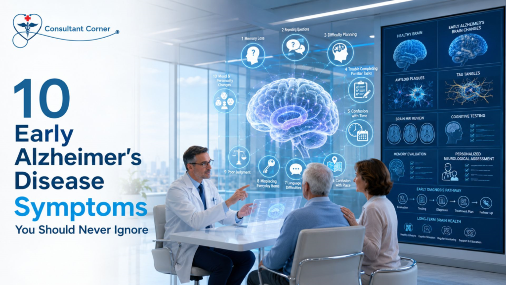
10 Early Alzheimer’s Disease Symptoms You Should Never Ignore Alzheimer’s Disease rarely announces itself all at once. It usually begins as something small — a missed appointment, a repeated question, a familiar word that suddenly feels out of reach. Because these moments look so much like ordinary forgetfulness, families often wait months or even years before asking a doctor whether something more serious might be going on. That delay matters. Recognizing Alzheimer’s Disease in its earliest stage gives patients and families more time to plan, more treatment options to consider, and a much better chance of protecting independence for as long as possible. At Consultant Corner, our neurology team evaluates memory concerns every week, and one pattern shows up again and again: the people who come in early almost always have more options than the people who wait. Below are the 10 early Alzheimer’s Disease symptoms our neurologists want every patient and caregiver to know, along with what to do if you recognize them in yourself or someone you love. It’s worth repeating that Alzheimer’s Disease is not a single dramatic event. It’s a gradual disease process, and the symptoms below can appear subtly at first — noticed only by a spouse, an adult child, or a close friend who sees the person regularly. That’s exactly why this list exists: not to cause alarm over every forgotten name, but to help you recognize the difference between ordinary aging and a pattern that deserves a professional evaluation. Why Early Symptoms of Alzheimer’s Disease Are So Easy to Miss Alzheimer’s Disease develops gradually, and the brain changes associated with it — the buildup of amyloid plaques and tau tangles — can begin a decade or more before obvious symptoms appear. In those early years, memory lapses are often chalked up to stress, poor sleep, or “just getting older.” The problem is that Alzheimer’s Disease symptoms don’t stay the same size. What starts as an occasionally misplaced set of keys can, over time, progress into disorientation in familiar places or trouble managing medications and finances. The goal isn’t to panic over every forgotten name. It’s to notice a pattern — memory or thinking changes that are frequent, progressive, and start to interfere with daily life. That pattern is the signal worth paying attention to. 1. Memory Loss That Disrupts Daily Life This is the hallmark early symptom of Alzheimer’s Disease. It looks like forgetting recently learned information, missing important dates, asking the same question multiple times in a single conversation, or relying heavily on notes and reminders for things that used to be automatic. Occasionally forgetting a name and remembering it later is normal aging. Forgetting the conversation ever happened at all is different. 2. Difficulty Planning or Solving Problems People in the early stages of Alzheimer’s Disease often struggle to follow a recipe, manage monthly bills, or keep track of a familiar process at work. Tasks that involve numbers, sequencing, or concentration take noticeably longer and require more effort than they used to. 3. Trouble Completing Familiar Tasks Driving to a well-known location, using household appliances, or remembering the rules of a favorite game can suddenly become confusing. This is different from occasionally needing help figuring out a new gadget — it’s difficulty with tasks the person has done comfortably for years. 4. Confusion With Time or Place Losing track of dates, seasons, or the passage of time is a common early Alzheimer’s Disease symptom. Someone may forget where they are, how they got there, or struggle to understand something unless it’s happening in the present moment. 5. Vision and Spatial Problems Some people with Alzheimer’s Disease develop difficulty reading, judging distances, or determining color and contrast — which can also affect driving safety. These changes are sometimes mistaken for ordinary vision decline, so an evaluation should rule out both eye conditions and neurological causes. 6. New Problems With Words in Speaking or Writing Struggling to find the right word, stopping mid-sentence, repeating phrases, or calling familiar objects by the wrong name are all early warning signs. This goes beyond the occasional “it’s on the tip of my tongue” moment everyone experiences. 7. Misplacing Items and Losing the Ability to Retrace Steps Putting things in unusual places — like keys in the refrigerator — and being unable to retrace steps to find them is a recognized early symptom of Alzheimer’s Disease. It’s often accompanied by suspicion that others have taken or moved the item. 8. Decreased or Poor Judgment Alzheimer’s Disease can affect a person’s ability to make sound decisions, whether that means falling for scams, giving away large sums of money, or paying less attention to grooming and personal hygiene than usual. 9. Withdrawal From Work or Social Activities People experiencing early memory or cognitive changes may pull back from hobbies, social events, or work projects because they’re having trouble keeping up — and because they feel self-conscious about it. This withdrawal is often one of the first things family members notice. 10. Changes in Mood and Personality Confusion, suspicion, depression, fear, and anxiety are common as Alzheimer’s Disease progresses. A person may become easily upset at home, with friends, or when taken out of a comfortable, familiar routine. Risk Factors That Increase the Chance of Alzheimer’s Disease Not everyone develops Alzheimer’s Disease, and researchers are still working to fully understand why some people are affected and others are not. That said, several risk factors are well established: Age — the single biggest known risk factor; risk rises significantly after age 65. Family history and genetics — having a parent or sibling with Alzheimer’s Disease raises personal risk, though it doesn’t guarantee a diagnosis. Cardiovascular health — high blood pressure, high cholesterol, diabetes, and obesity are all linked to higher dementia risk, likely because they affect blood flow to the brain. Head injury history — a prior head injury or concussion, especially repeated trauma, is associated with increased long-term risk. Sleep quality — chronic poor sleep or untreated sleep apnea
11 Migraine Triggers That Could Be Causing Your Headaches
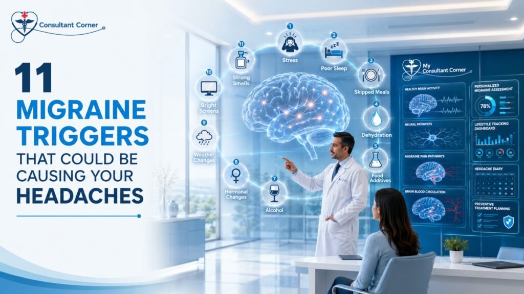
11 Migraine Triggers That Could Be Causing Your Headaches Headache & Migraine pain rarely shows up out of nowhere. For most people living with recurring attacks, an identifiable trigger — or a combination of several — sets the pain in motion hours or even a full day before the first throb hits. If you have ever wondered why one week brings three migraines and the next brings none, the answer usually isn’t bad luck. It’s a pattern hiding in your sleep schedule, your diet, your stress levels, or your environment, and once you can name it, you can start managing it. At Consultant Corner, we evaluate Headache & Migraine patients every week who are surprised to learn just how many everyday factors can set off an attack. Some triggers are obvious, like a stressful deadline or a missed meal. Others are far more subtle — a shift in barometric pressure, a whiff of perfume, or an extra hour of sleep on a Saturday morning. Because migraine triggers rarely act alone, most patients are dealing with a combination of two or three factors stacking together before an attack finally breaks through. Below, we break down the 11 most common migraine triggers, explain the science behind each one, and share practical steps you can take to reduce how often — and how severely — migraines interrupt your life. Whether you experience an occasional migraine or you’re managing a chronic pattern of frequent attacks, understanding what’s setting off your pain is the foundation of any effective treatment plan. Why Identifying Your Headache & Migraine Triggers Matters Migraine is a neurological condition, not “just a bad headache.” Research from the Mayo Clinic explains that migraine attacks can cause intense throbbing pain, nausea, and extreme sensitivity to light and sound, and they can last anywhere from a few hours to several days. Because the brain of someone with migraine disease is more sensitive to change than the average brain, small shifts — a missed meal, a poor night’s sleep, a change in the weather — can be enough to trigger a full attack. That sensitivity is exactly why trigger identification matters so much. Our team at Consultant Corner walks patients through the process of connecting the dots between lifestyle patterns and headache frequency, which is also covered in more depth on our Understanding Headache / Migraine page. A headache and migraine diary — tracking sleep, meals, stress, weather, and menstrual cycle alongside every attack — is often the single most useful tool a patient can bring to their first appointment. It’s also worth understanding that triggers and causes are not the same thing. Migraine is a genetic, neurological condition — you don’t develop it because of stress or bad habits. Triggers simply lower the threshold your brain needs to start an attack on a day when your nervous system is already primed to react. That’s why the same trigger, say a glass of red wine, might cause a migraine one week and nothing at all the next: it depends on what else is happening in your body at the time. Recognizing this interplay helps patients stop blaming themselves for “doing something wrong” and instead focus on the practical work of pattern-tracking and prevention. Below are the 11 triggers we see most often in our Headache & Migraine clinic, along with what the research says about why they matter. The 11 Most Common Migraine Triggers 1. Stress and Emotional Tension Stress is consistently ranked as the single most reported migraine trigger. When the body is under sustained pressure, it releases hormones like cortisol that can affect blood vessels and nerve signaling in the brain. Interestingly, it isn’t only stress itself that causes problems — the sudden drop in stress after a demanding week (sometimes called a “let-down migraine”) can be just as provocative. Simple stress-reduction techniques, including breathing exercises, short walks, and consistent downtime, can meaningfully reduce Headache & Migraine frequency over time. 2. Irregular Sleep Patterns Both too little sleep and too much sleep can trigger a migraine. Migraine sufferers tend to have brains that crave consistency, so going to bed and waking up at wildly different times — even on weekends — can be enough to set off an attack. According to the American Migraine Foundation, sleeping too much or too little is one of the most common triggers reported by patients. Aiming for a steady 7–8 hours each night is one of the simplest, most effective preventive habits available. 3. Skipped Meals and Dehydration Skipping meals causes blood sugar to drop, which can trigger a migraine in susceptible individuals. Dehydration works the same way — even mild fluid loss can reduce blood flow to the brain and bring on head pain. Keeping regular mealtimes and carrying water throughout the day are two of the easiest changes a Headache & Migraine patient can make. 4. Certain Foods and Food Additives Aged cheeses, processed meats containing nitrates, foods with monosodium glutamate (MSG), and artificial sweeteners are frequently reported migraine triggers. Not every migraine patient reacts to the same foods, which is why a food-and-headache journal is so valuable — it helps identify your personal pattern rather than relying on a generic “avoid this list.” 5. Caffeine and Alcohol Caffeine is a double-edged sword. A small amount can actually relieve an early migraine, but too much — or sudden caffeine withdrawal on a weekend when your routine changes — can trigger one. Alcohol, especially red wine and other beverages containing sulfites and histamines, is another well-documented trigger. Moderation and consistency (rather than complete avoidance) tend to work best for most patients. 6. Hormonal Changes Migraine affects women roughly three times more often than men, largely because of fluctuations in estrogen. Many women notice a strong connection between their menstrual cycle and Headache & Migraine attacks, with pain often striking in the days just before or during menstruation. Pregnancy, perimenopause, and hormonal medications like oral contraceptives can also shift migraine frequency and severity. 7. Weather and
12 Early Ischemic Stroke Symptoms That Could Save Your Life

12 Early Ischemic Stroke Symptoms That Could Save Your Life These are the warning signs of an ischemic stroke that deserve immediate attention. Some appear together, and some show up in isolation, but any single one of them is enough reason to call 911. 1. Sudden Numbness or Weakness on One Side The most classic sign of an ischemic stroke is a sudden loss of feeling or strength that affects only one side of the body — often the face, arm, or leg. This happens because each side of the brain controls the opposite side of the body, so a blockage on one side produces symptoms on the other. 2. Facial Drooping One side of the face may suddenly droop, feel numb, or fail to move normally when the person tries to smile. Facial drooping is one of the three checks used in the FAST test and is often the most visible sign to bystanders. 3. Arm Weakness or Drift Ask the person to raise both arms. If one arm drifts downward or cannot be lifted at all, this points toward a stroke affecting the motor pathways on one side of the brain. 4. Slurred or Garbled Speech Sudden difficulty forming words, slurring, or speech that sounds unusually thick or slow can indicate that an ischemic stroke is affecting the areas of the brain responsible for language and motor coordination of the mouth and tongue. 5. Trouble Understanding Others Some people having an ischemic stroke can speak but cannot understand what’s being said to them, or they respond with words that don’t make sense. This is a sign that a language-processing region of the brain has been affected. 6. Sudden Vision Loss or Blurriness Vision changes, including blurred vision, double vision, or a sudden loss of sight in one or both eyes, are common but frequently overlooked symptoms of an ischemic stroke, especially when they aren’t paired with obvious weakness. 7. Sudden, Severe Headache A headache that comes on abruptly and feels unlike any prior headache — often described as the “worst headache of my life” — should never be dismissed. While this is more classically linked to hemorrhagic stroke, it can also accompany certain ischemic events. 8. Dizziness or Loss of Balance A sudden inability to walk steadily, an unexplained loss of coordination, or a feeling that the room is spinning can be an early sign of an ischemic stroke affecting the brainstem or cerebellum. For a deeper look at causes of dizziness beyond stroke, see Vertigo & Dizziness: 9 Common Causes. 9. Sudden Confusion A sudden change in mental clarity — becoming disoriented, forgetting where you are, or struggling to follow a simple conversation — can be an easily missed symptom of an ischemic stroke, particularly in older adults. 10. Difficulty Walking Beyond dizziness, an ischemic stroke can cause a sudden stumble, a dragging leg, or an inability to coordinate the steps needed to walk normally, even without any pain. 11. Numbness or Tingling That Comes and Goes Symptoms that appear and then resolve within minutes can still represent a serious warning. This pattern is often the hallmark of a transient ischemic attack, a short-lived event that frequently precedes a full ischemic stroke. 12. Sudden Nausea or Vomiting With Other Symptoms On its own, nausea isn’t a stroke symptom, but when it appears alongside any of the signs above — especially dizziness, confusion, or weakness — it should raise immediate concern for an ischemic stroke rather than being written off as a stomach bug. Use the FAST Test to Recognize an Ischemic Stroke The FAST test remains the fastest, most reliable way to identify an ischemic stroke in the moment, and it doesn’t require any medical training to use. Letter What to Check What It Means F – Face Ask the person to smile Does one side of the face droop? A – Arms Ask the person to raise both arms Does one arm drift downward or feel weak? S – Speech Ask the person to repeat a simple sentence Is speech slurred, strange, or hard to understand? T – Time Note when symptoms started Call 911 immediately — do not wait or drive yourself Some hospitals now teach an expanded version, BE-FAST, which adds Balance and Eyes to catch strokes affecting coordination or vision. According to the American Stroke Association, learning and sharing the B.E. F.A.S.T. warning signs of stroke could help save a life. Either version works — what matters is acting the moment any of these checks come back abnormal. Why Fast Action Against an Ischemic Stroke Saves Lives Treatment for an ischemic stroke is highly time-dependent. When someone arrives at the hospital quickly enough, physicians may be able to use a clot-dissolving medication or a mechanical procedure to physically remove the blockage, both of which are far more effective the sooner they’re administered. The CDC notes that patients with ischemic strokes who receive clot-busting medication are more likely to recover fully or have less disability than patients who do not receive the drug, and are also less likely to need long-term care in a nursing home. This is also why a transient ischemic attack, or TIA, should never be brushed off just because the symptoms resolved on their own. A TIA is essentially a preview of a future ischemic stroke, and ignoring it removes the opportunity to intervene before a larger, more damaging event occurs. If you’ve experienced symptoms that came and went, it’s worth reading our full breakdown of TIA warning signs to understand how closely the two conditions are connected. Ischemic Stroke vs. Hemorrhagic Stroke Not all strokes are caused by a blockage. Understanding the difference helps explain why treatment approaches vary so much between the two. Feature Ischemic Stroke Hemorrhagic Stroke Cause Blocked artery (clot or plaque) Ruptured blood vessel Frequency About 87% of all strokes About 13% of all strokes Common Treatment Clot-dissolving medication, clot removal Surgery, blood pressure control Key Risk Factors High blood pressure, high cholesterol, atrial
10 Warning Signs of a Head Injury You Should Never Ignore
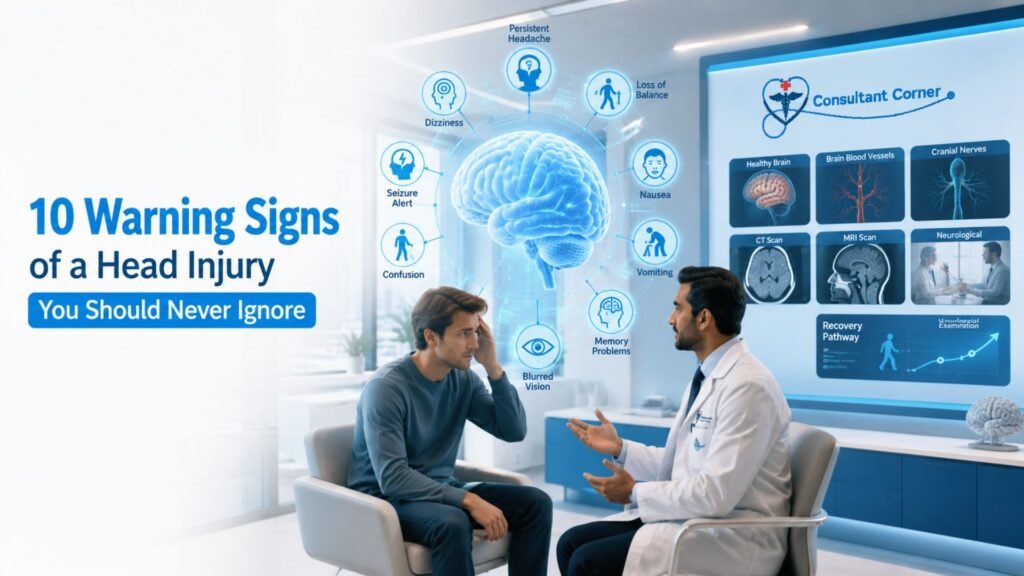
10 Warning Signs of a Head Injury You Should Never Ignore A head injury can look harmless one moment and turn serious the next. You bump your head getting out of the car, you take a fall on the stairs, or you’re in a minor fender-bender — and you feel fine. Then, hours or even days later, a headache creeps in, your thinking feels foggy, or the room starts to spin. This delayed pattern is exactly why this kind of injury is so easy to underestimate and so important to take seriously. At Consultant Corner, our neurology team evaluates patients with a head injury every week, from athletes and weekend warriors to older adults who’ve had a simple fall at home. Below, we walk through the 10 warning signs you should never ignore, explain why each one matters, and outline exactly when a bump on the head needs a same-week neurology visit — or an emergency room. Why a Head Injury Deserves Immediate Attention Every head injury is different because every brain responds to trauma differently. A blow to the head can range from a mild bump that resolves on its own to a traumatic brain injury (TBI) that causes swelling, bleeding, or lasting damage. According to the Centers for Disease Control and Prevention, some signs of a serious brain injury appear immediately, while others don’t show up until hours or days later — which is exactly why “I feel okay right now” isn’t the same as “I’m in the clear.” The danger with this kind of trauma is that the brain is a soft, delicate organ cushioned inside a hard skull. A sudden jolt, blow, or rapid deceleration can cause the brain to move within the skull, stretching nerve fibers and, in more serious cases, causing bruising or bleeding. That’s why doctors urge anyone who has experienced this kind of trauma — even a “minor” bump — to watch closely for the warning signs described below. Common Causes of a Head Injury Understanding how these injuries typically happen can help you recognize your own risk level. The most frequent causes include: Falls — the leading cause across all age groups, especially among young children and older adults Motor vehicle collisions — including car, motorcycle, and bicycle accidents Sports and recreational activities — contact sports like football, hockey, and soccer, as well as cycling and skiing Workplace accidents — falling objects, equipment accidents, or falls from height Physical altercations or assaults Regardless of the cause, the same principle applies: any forceful blow, jolt, or sudden stop that moves the brain inside the skull has the potential to cause lasting harm, so the mechanism of injury matters less than how closely you monitor your symptoms afterward. How to Reduce Your Risk of a Head Injury While not every accident can be prevented, several everyday habits meaningfully lower your risk: Wear a properly fitted helmet for biking, skiing, contact sports, and motorcycle riding Use seatbelts and appropriate child car seats on every trip, even short ones Remove fall hazards at home, such as loose rugs, poor lighting, and cluttered stairways Install handrails and grab bars for older adults or anyone with balance concerns Follow return-to-play guidelines after a prior concussion instead of rushing back into sports These small precautions won’t eliminate risk entirely, but they meaningfully reduce both the likelihood and severity of trauma to the head. Children, Older Adults, and Head Injury Risk Two groups deserve extra caution after any blow to the head: young children and older adults. Kids may not be able to describe symptoms like fogginess or light sensitivity, so parents should watch for excessive crying, unusual sleepiness, or refusal to eat or nurse. Older adults, especially those taking blood thinners, face a higher risk of bleeding inside the skull even after a seemingly minor fall, and symptoms can be subtler — mistaken for normal fatigue or confusion rather than a medical emergency. If a child or an older family member has taken a fall or a hit to the head, it’s worth erring on the side of caution and scheduling an evaluation, even if they seem fine at first. 10 Warning Signs of a Head Injury You Should Never Ignore 1. A Headache That Won’t Go Away or Keeps Getting Worse A dull ache after a bump is common, but a headache that intensifies over hours, rather than fading, is one of the most important warning signs on this list. A worsening headache can point to swelling or bleeding building up pressure inside the skull. This is different from a typical tension headache, which usually stays mild and steady rather than escalating. 2. Dizziness, Loss of Balance, or Feeling “Off” Struggling to walk a straight line, feeling unsteady on your feet, or sensing that something is generally “off” after trauma to the head often signals that the impact has disrupted the brain pathways responsible for coordination and spatial awareness. This can appear right away or develop over the following day. 3. Nausea or Repeated Vomiting Occasional queasiness after a jolt to the head isn’t unusual, but nausea that persists — or vomiting more than once — is a recognized sign of increased pressure inside the brain and should never be brushed off, especially in children and older adults. 4. Trouble Concentrating, Remembering, or Processing Information Many people describe this as feeling “foggy,” mentally slow, or unable to hold a thought after a blow to the head. Difficulty concentrating at work, forgetting recent conversations, or struggling to follow a simple task are classic post-concussion symptoms that deserve a proper neurological evaluation. 5. Sensitivity to Light or Blurry Vision Photophobia (light sensitivity) and visual disturbances such as blurred or double vision can appear immediately or emerge days after the trauma. These symptoms suggest the brain is still irritated from the impact and hasn’t finished healing. 6. Sudden, Severe Headache Unlike Any Before A “thunderclap” headache — one that comes on suddenly and feels far more intense than any
10 Recovery Tips to Avoid Post-Stroke Setbacks
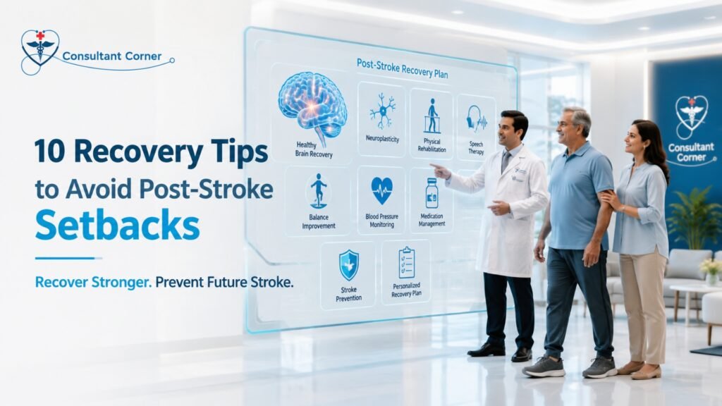
10 Recovery Tips to Avoid Post-Stroke Setbacks Post-Stroke recovery doesn’t end when you leave the hospital — it’s a daily process, and small mistakes in those first weeks and months can quietly undo the progress you’ve worked hard to make. Whether you or someone you love is a few days or several months into healing, understanding how to protect that progress is one of the most important things you can do. Below, we walk through ten practical, medically informed tips that can help you avoid the most common post-stroke setbacks and keep recovery moving in the right direction. A stroke is a sudden, life-altering event, but what happens after the hospital discharge often determines the long-term outcome. Weakness, coordination issues, balance problems, memory changes, fatigue, speech difficulty, and emotional swings can all persist for weeks, months, or years. The encouraging news is that with the right follow-up care, structured rehabilitation, and a few key lifestyle adjustments, most patients recover faster, stronger, and safer than they expect. At Consultant Corner, our neurology team has guided hundreds of patients through this critical recovery window, and we’ve seen firsthand which habits accelerate healing — and which ones quietly cause setbacks. This guide brings those lessons together in one place. No two recoveries look exactly alike, and comparing your own timeline to someone else’s story online can do more harm than good. Some patients regain independence within a few months, while others need a year or more of structured therapy before they feel like themselves again. What almost every successful recovery has in common, though, is a consistent, well-coordinated care plan — one that treats the body, the brain, and the emotional side of healing as equally important. That’s the philosophy behind every tip below. Why the Post-Stroke Recovery Window Matters So Much The brain continues to heal and rewire itself for 12 to 24 months after a stroke, a process doctors call neuroplasticity. During this period, the brain is unusually responsive to therapy, repetition, and structured stimulation. Without proper guidance, however, patients often experience slow recovery, persistent weakness, a higher risk of falls, unmanaged spasticity, and — in the most serious cases — a repeat stroke. This is exactly why Post-Stroke follow-up care isn’t optional; it’s the foundation that everything else is built on. According to the Centers for Disease Control and Prevention, recovery time after a stroke differs for everyone and can take weeks, months, or even years, with some people recovering fully while others face long-term or lifelong disabilities. That variability is precisely why an individualized recovery plan — not a generic checklist — matters so much. 10 Recovery Tips to Avoid Post-Stroke Setbacks 1. Never Skip Your Neurology Follow-Up Appointments It’s tempting to feel like you’re “done” with medical care once you’re home, but Post-Stroke follow-up visits are where your care team catches subtle changes before they become major setbacks. Regular evaluation of strength, balance, speech, and cognition allows your neurologist to adjust medications, therapy intensity, and safety recommendations as your brain heals. Patients who skip these visits are far more likely to experience preventable complications, from unmanaged spasticity to missed signs of a second stroke. 2. Stick to Your Rehabilitation Schedule — Even on Hard Days Physical, occupational, and speech therapy are not optional extras; they are the engine of Post-Stroke recovery. The brain rebuilds neural pathways through repetition, so missing sessions — even a few — can slow momentum that took weeks to build. On days when fatigue or frustration makes therapy feel impossible, talk to your care team about modifying the session rather than skipping it entirely. Consistency, not intensity, is what drives long-term gains. 3. Manage Blood Pressure, Cholesterol, and Blood Sugar Closely One of the most common causes of a second stroke is poorly controlled cardiovascular risk factors. High blood pressure, elevated cholesterol, and unmanaged diabetes all place ongoing strain on the blood vessels that supply the brain. Home monitoring, consistent medication use, and regular lab work are simple habits that meaningfully lower the risk of another event during Post-Stroke recovery. It helps to think of these numbers as an early warning system rather than an inconvenience. A home blood pressure cuff, a simple daily log, and a shared spreadsheet or notebook with your care team can catch a dangerous trend weeks before it becomes an emergency. Diet also plays a bigger role than most patients expect — reducing sodium, limiting processed foods, and increasing fiber and lean protein intake can measurably improve blood pressure and cholesterol numbers within a few months. Ask your neurologist or a referred dietitian for a plan tailored to your specific risk factors, since generic advice rarely accounts for medication interactions or kidney function. 4. Address Swallowing and Speech Difficulties Early Speech and swallowing problems (dysphagia) are common after a stroke and are often underreported by patients who feel embarrassed or assume the issue will resolve on its own. Left untreated, swallowing difficulties can lead to choking, aspiration pneumonia, and malnutrition — all of which can dramatically derail recovery. A speech-language pathologist can assess swallowing safety and provide targeted exercises that improve both speech clarity and swallowing function over time. 5. Reduce Fall Risk at Home Balance and coordination problems make falls one of the most frequent post-stroke setbacks, and a single fall can result in a fracture or head injury that resets months of progress. Simple home modifications — removing loose rugs, installing grab bars in the bathroom, improving lighting, and using assistive devices as recommended — go a long way. If dizziness or vertigo has been part of your recovery, it’s worth discussing with your care team, since balance issues sometimes overlap with other neurological conditions. A physical or occupational therapist can also perform a formal fall-risk assessment, walking through your home environment room by room to flag specific hazards you might not notice day to day — a slick bathroom floor, a poorly lit hallway, or stairs without a secure handrail. Footwear matters too; loose slippers
8 Muscle Weakness Signs That May Need ALS Screening
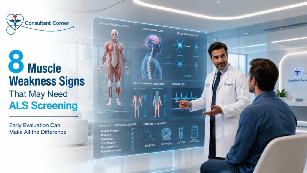
8 Muscle Weakness Signs That May Need ALS Screening Feeling weaker than usual? Losing strength in your arms, legs, or hands? Don’t ignore it — early evaluation can make all the difference. Muscle weakness is one of the most common reasons patients schedule a neurology appointment, and in the overwhelming majority of cases, it is caused by something treatable — a pinched nerve, a vitamin deficiency, thyroid dysfunction, or a compressed disc. But when muscle weakness becomes progressive, spreads from one area of the body to another, or shows up alongside twitching and unexplained fatigue, it is important to rule out rarer but more serious neurological causes, including amyotrophic lateral sclerosis (ALS). At Consultant Corner, muscle weakness and ALS screening are evaluated with the same level of urgency and precision. Our goal is simple: identify the true cause of your weakness quickly, rule out what it isn’t, and build a clear plan forward — whether that means treating a reversible condition or connecting you with the right specialists for further workup. This guide walks through eight muscle weakness signs that deserve a closer look, explains what typically causes muscle weakness, and outlines what a thorough ALS screening actually involves. Why Muscle Weakness & ALS Screening Matters Most people who notice new weakness assume the worst, and that fear alone can keep people from seeking care. The truth is reassuring: the vast majority of muscle weakness has nothing to do with ALS. Nerve compression, neuropathy, and metabolic issues account for the majority of weakness complaints seen in neurology clinics. Still, because ALS is a progressive disease where early diagnosis and early treatment meaningfully affect outcomes, muscle weakness and ALS screening should never be delayed when the warning signs listed below are present. Screening does two things at once. First, it identifies and treats the common, reversible causes of weakness that are affecting most patients. Second, it catches the small subset of cases where early neurological disease is present, so treatment can begin as soon as possible. 8 Warning Signs of Muscle Weakness That Warrant Evaluation If you’re noticing any of the following, it’s worth scheduling a muscle weakness and ALS screening evaluation rather than waiting to see if it improves on its own. 1. One Arm or Leg Becoming Progressively Weaker Weakness that is limited to one side of the body — and that continues to get worse over weeks or months rather than staying stable — is one of the more notable patterns doctors look for. Weakness that stays the same for months and doesn’t spread is far less concerning than weakness that keeps advancing. 2. Difficulty Lifting Objects or Gripping Items Trouble opening jars, holding a coffee cup, or gripping a steering wheel can point to problems ranging from carpal tunnel syndrome to cervical radiculopathy to early motor neuron changes. A hand-strength decline that shows up on both sides equally is usually different — and typically less urgent — than weakness isolated to one hand. 3. Foot Drop or Frequent Tripping Foot drop happens when the muscles that lift the front of the foot weaken, causing the toes to catch on the ground while walking. It’s a hallmark of peroneal nerve compression, but it can also be an early sign of more diffuse motor neuron involvement, which is why it’s included in a thorough muscle weakness and ALS screening. 4. Trouble Raising Your Arms or Climbing Stairs Weakness that affects the muscles closest to the body — shoulders, hips, and thighs — rather than the hands and feet has a different pattern of causes, including myopathies and certain autoimmune muscle diseases. Struggling to reach overhead or push up from a chair are both signals worth mentioning to your doctor. 5. Muscle Twitching (Fasciculations) Occasional muscle twitches, especially in the calves or eyelids, are extremely common and are almost always benign, often related to caffeine, stress, dehydration, or fatigue. Twitching that is widespread, persistent, and paired with muscle weakness is the combination that warrants a closer look during ALS screening — twitching alone, without accompanying weakness, is rarely a red flag. 6. Unexplained Fatigue With Simple Tasks Feeling unusually exhausted after routine activities — climbing a single flight of stairs, carrying groceries, or brushing your hair — can reflect anything from anemia and thyroid disease to early neuromuscular weakness. Because fatigue overlaps with so many common conditions, it’s one of the harder symptoms to interpret without proper testing. 7. Handwriting Becoming Smaller or Shakier A noticeable change in handwriting — letters shrinking, lines becoming less steady, or grip fatigue mid-sentence — can reflect fine motor weakness in the hand. While this symptom is also associated with Parkinson’s disease, in the context of progressive limb weakness it belongs on the muscle weakness and ALS screening checklist. 8. Slurred Speech, Trouble Swallowing, or Voice Changes When weakness begins to affect the muscles used for speaking or swallowing — a nasal-sounding voice, slurred words, or choking on liquids — this is one of the symptoms that should prompt an immediate evaluation, since it can also signal a stroke in its acute form. Any sudden change here deserves urgent attention. What Causes Muscle Weakness? (Most of It Isn’t ALS) It bears repeating: most muscle weakness is not ALS. A thorough muscle weakness and ALS screening looks at the full range of possible causes, including: Pinched nerves and nerve entrapment (such as carpal tunnel syndrome) Peripheral neuropathy Cervical or lumbar radiculopathy (nerve root compression in the neck or lower back) Stroke or transient ischemic attack (TIA)-related weakness Myopathies (primary muscle diseases) Thyroid disorders, including hypothyroidism Vitamin deficiencies, particularly B12 and vitamin D Autoimmune neuromuscular conditions, such as myasthenia gravis Medication side effects, including certain statins and steroids Because so many of these conditions overlap in how they present, an evaluation for muscle weakness and ALS screening is designed to systematically rule each of them in or out — not jump straight to worst-case scenarios. Who Is at Higher Risk for ALS? Understanding risk factors
9 Sleep Disorders & Restless Legs Syndrome (RLS) Signs You Shouldn’t Ignore
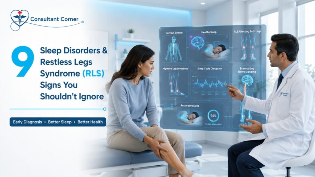
9 Sleep Disorders & Restless Legs Syndrome (RLS) Signs You Shouldn’t Ignore Restless Legs Syndrome (RLS) is one of the most overlooked reasons people spend years exhausted, irritable, and unable to explain why a full eight hours in bed never seems to leave them rested. If you’ve been tossing, turning, kicking off the blankets, or feeling a strange crawling sensation in your legs the moment you finally lie down, you are dealing with something real — and treatable. Sleep disorders and Restless Legs Syndrome (RLS) don’t just steal your nights; they quietly erode your mood, your focus at work, your relationships, and your long-term health. At Consultant Corner, our neurology team evaluates sleep disorders and Restless Legs Syndrome (RLS) every week, and the pattern is almost always the same: patients assume their symptoms are “just stress” or “just getting older” until the disruption becomes impossible to ignore. Below are nine warning signs that your sleep trouble may be rooted in a diagnosable neurological condition — and what you can do about it. What Are Sleep Disorders and Restless Legs Syndrome (RLS)? Sleep disorders are a broad category of conditions that interfere with your ability to fall asleep, stay asleep, or feel rested after sleep. According to the National Heart, Lung, and Blood Institute, tens of millions of Americans live with a diagnosable sleep disorder, and many never receive a formal diagnosis or treatment. Restless Legs Syndrome (RLS) is a specific neurological movement disorder that sits within this larger category. It causes an uncomfortable, sometimes indescribable urge to move the legs, usually in the evening or at night, and usually only relieved by movement. As Mayo Clinic explains, this nightly urge to move can interrupt sleep so consistently that people end up dragging through their entire day — and for many patients, that daytime exhaustion is the symptom that finally sends them looking for answers. Sleep issues can also follow a head injury or concussion, a connection our team explores in an earlier post, What is a Head Injury? Because Restless Legs Syndrome (RLS) frequently overlaps with insomnia, periodic limb movement disorder, and other neurological conditions, an accurate diagnosis requires more than guesswork. It requires a clinician who understands how these conditions interact. 9 Signs of Sleep Disorders & Restless Legs Syndrome (RLS) You Shouldn’t Ignore 1. Trouble Falling Asleep or Staying Asleep If it regularly takes you 30 minutes or longer to fall asleep, or you wake up multiple times a night and struggle to drift back off, this isn’t simply “a bad sleeper’s habit.” Chronic difficulty initiating or maintaining sleep is one of the defining features of insomnia, and when it’s paired with leg discomfort, it often points toward Restless Legs Syndrome (RLS) as the underlying driver. 2. An Uncomfortable “Crawling” or “Pulling” Sensation in the Legs People describe this sensation in strikingly consistent terms — creeping, tingling, throbbing, aching, electric, or itchy deep inside the muscle rather than on the skin. It typically shows up in the calves or thighs, though it can occasionally affect the arms. This sensation is one of the clearest diagnostic markers of Restless Legs Syndrome (RLS), and it’s rarely caused by anything other than a neurological condition. 3. An Urge to Move Your Legs, Especially at Night The hallmark symptom of Restless Legs Syndrome (RLS) isn’t just discomfort — it’s an almost irresistible urge to move. Patients describe needing to shake, stretch, pace, or flex their legs to get any relief. This urge tends to intensify the longer you sit or lie still, which is why it so often strikes right as you’re trying to fall asleep. 4. Symptoms That Worsen in the Evening or at Night A distinguishing feature of Restless Legs Syndrome (RLS) is its circadian pattern: symptoms are typically mild or absent during the day and become noticeably worse in the evening and overnight. If your legs feel fine at your desk but start acting up the moment you sit down for dinner or climb into bed, that timing pattern is medically significant. 5. Involuntary Leg Twitching or Jerking During Sleep Many people with Restless Legs Syndrome (RLS) also experience periodic limb movements of sleep — rhythmic kicking or jerking that occurs every 20 to 40 seconds throughout the night, often without the person waking up or even remembering it. A bed partner is frequently the first to notice this pattern, and it can fragment sleep just as severely as full waking episodes. 6. Frequent Nighttime Waking, Vivid Dreams, or Confusion Fragmented sleep architecture — the repeated cycling in and out of light sleep — often produces unusually vivid or unsettling dreams, along with brief moments of grogginess or confusion upon waking. When this happens night after night, it’s a sign your sleep cycle isn’t completing its normal restorative stages. 7. Daytime Fatigue, Brain Fog, and Difficulty Concentrating Perhaps the most disruptive consequence of sleep disorders and Restless Legs Syndrome (RLS) is what happens during the day. Patients often describe needing extra caffeine just to function, struggling to focus in meetings, forgetting simple tasks, or feeling irritable for no clear reason. This daytime impairment is frequently what finally prompts someone to seek a specialist. 8. Symptoms That Improve With Movement but Return With Rest Because Restless Legs Syndrome (RLS) symptoms are relieved — even temporarily — by walking, stretching, or shifting position, many patients develop unconscious coping habits: pacing before bed, sleeping with a leg outside the covers, or getting up repeatedly through the night. If you find yourself needing to move just to get comfortable, that relief-with-movement pattern is a key diagnostic clue. 9. Family History of Restless Legs Syndrome (RLS) or Unexplained Sleep Trouble Restless Legs Syndrome (RLS) has a strong genetic component, particularly in cases that begin before age 45. If a parent or sibling has been diagnosed with RLS, or has described similar “can’t keep my legs still” symptoms, your own leg discomfort deserves a proper neurological evaluation rather than being dismissed as unrelated. What
7 TIA (Mini-Stroke) Warning Signs You Should Learn
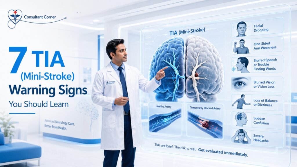
7 TIA (Mini-Stroke) Warning Signs You Should Learn TIA warning signs are easy to miss because they vanish almost as quickly as they appear — and that is exactly what makes them so dangerous. A sudden change in vision, a wave of weakness down one arm, or words that suddenly won’t come out right can resolve in minutes and leave a person wondering if anything really happened at all. It did. Recognizing TIA warning signs the moment they occur, rather than brushing them off once they pass, is one of the most important things you can do to protect your brain from a future stroke. A Transient Ischemic Attack (TIA), often called a “mini-stroke,” is one of the most important warning signs in neurology. Symptoms may last only minutes, but the risk behind them is very real. Research consistently shows that a meaningful share of people who experience a TIA go on to have a major stroke, and a large portion of those strokes happen within the first two days afterward. That narrow window is why urgent evaluation — not a “wait and see” approach — is critical. At Consultant Corner, we specialize in rapid neurological assessment after a TIA to help prevent a life-changing stroke. This guide walks through the seven TIA warning signs you should learn to recognize, why TIAs happen, what causes them, and what to do the moment symptoms appear — even if they’ve already gone away by the time you read this. What Is a TIA (Mini-Stroke)? A TIA occurs when blood flow to part of the brain is briefly interrupted, usually by a small clot that lodges in an artery and then breaks apart or dislodges on its own. Unlike a full ischemic stroke, a TIA does not cause permanent tissue damage, which is why symptoms typically resolve within minutes to an hour. But the underlying problem — the clot, the narrowed artery, the irregular heart rhythm, or the uncontrolled blood pressure — is still there after the symptoms fade. That’s the paradox of TIA warning signs: the body sends a clear signal, then appears to take it back. Many patients feel fine within the hour and decide not to seek care. This is one of the most common and most dangerous mistakes in stroke prevention, because a TIA is, in effect, the brain’s way of announcing that a bigger event may be coming. Understanding TIA Warning Signs Early Neurologists sometimes describe a TIA as a “warning stroke.” It behaves like a stroke, mimics the symptoms of a stroke, and shares the same underlying causes as a stroke — the only difference is duration. That single distinction is why so many people underestimate it. Why TIA Warning Signs Demand Urgent Attention The numbers behind TIA warning signs are sobering: Roughly 1 in 3 patients who experience a TIA will go on to have a major stroke. Up to 50% of those strokes occur within the first 48 hours after the TIA. Risk remains elevated for weeks afterward, which is why a structured evaluation and prevention plan matters as much as the initial visit. These figures explain why emergency medicine and neurology both treat TIA the same way they treat an active stroke: as a time-sensitive emergency, not a symptom to monitor at home. According to the National Institute of Neurological Disorders and Stroke (NINDS), TIA symptoms are similar to stroke symptoms and typically resolve within an hour, though they can occasionally last up to 24 hours — and there is no reliable way to tell, in the moment, whether you are having a TIA or a full stroke without medical evaluation. 7 TIA Warning Signs You Should Learn Below are the seven core TIA warning signs that should prompt an immediate call for help. Symptoms may last only a few minutes, but treat every one of these as an emergency, regardless of how quickly it resolves. 1. Sudden Weakness or Numbness on One Side of the Body This is one of the most recognizable TIA warning signs. It typically affects the face, arm, or leg on a single side of the body and comes on abruptly, without any obvious trigger. A drooping side of the face, a hand that suddenly can’t grip a cup, or a leg that buckles unexpectedly all fall into this category. 2. Slurred Speech or Trouble Finding Words Sudden difficulty speaking clearly, understanding what others are saying, or finding the right words mid-sentence is a hallmark neurological warning sign. It often appears alongside facial drooping and is one of the fastest ways bystanders can identify a possible TIA or stroke in progress. 3. Vision Loss, Double Vision, or Temporary Blindness in One Eye Sudden vision changes — blurring, dimming, double vision, or a curtain-like loss of sight in one eye — are frequently overlooked TIA warning signs because they’re often mistaken for eye strain or migraine aura. When vision changes appear suddenly and resolve just as quickly, they deserve the same urgency as any other stroke symptom. 4. Dizziness, Imbalance, or Sudden Inability to Walk A sudden loss of coordination, a feeling that the room is tilting, or an inability to walk a straight line can indicate that blood flow has been disrupted in the areas of the brain responsible for balance. This symptom is sometimes confused with inner-ear vertigo, which is why a proper neurological evaluation matters — read more about the overlap in our related post on Vertigo & Dizziness: 9 Common Causes. 5. Confusion or Difficulty Understanding Speech Sudden confusion, trouble following a conversation, or an inability to process simple instructions can be subtle but important TIA warning signs, particularly in older adults where confusion is sometimes mistakenly attributed to fatigue or age. 6. Sudden, Unexplained Headache A severe headache that appears out of nowhere, without a clear trigger, and feels different from a typical headache or migraine, can accompany a TIA. If you experience recurring headache patterns and want to understand the difference between a
Vertigo & Dizziness: 9 Common Causes
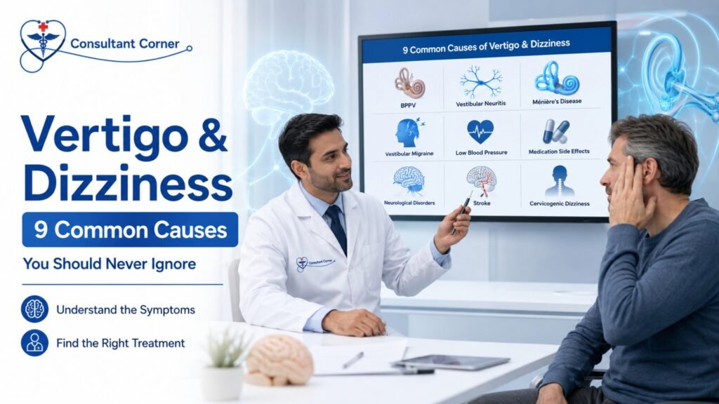
Vertigo & Dizziness: 9 Common Causes Vertigo and dizziness are two of the most common — and most confusing — symptoms a person can experience. One moment you’re standing in your kitchen, and the next the room feels like it’s tilting, spinning, or dropping out from under you. For many people, vertigo and dizziness show up suddenly, with no warning, and then vanish just as quickly, leaving behind a nagging fear that it will happen again. If you’ve been searching for answers about vertigo and dizziness, you’re not alone. Millions of adults experience these symptoms every year, and while an occasional dizzy spell after standing up too fast is usually harmless, recurring vertigo and dizziness are your body’s way of telling you that something in your inner ear, brain, or nervous system needs attention. At Consultant Corner, our neurology team specializes in identifying exactly what’s driving your symptoms and creating a plan to help you feel steady again. In this guide, we’ll walk through the nine most common causes of vertigo and dizziness, the warning signs that mean you should be evaluated right away, and what treatment typically looks like once a diagnosis is made. What’s the Difference Between Vertigo and Dizziness? Although people often use the words interchangeably, vertigo and dizziness aren’t quite the same thing. Vertigo refers specifically to the false sensation that you, or the room around you, is spinning or moving. Dizziness is a broader term that can describe lightheadedness, faintness, unsteadiness, or a floating sensation without any spinning at all. Understanding this distinction matters because it helps your provider narrow down the cause. Spinning sensations tend to point toward the inner ear or the brainstem, while lightheadedness more often points toward blood pressure, circulation, or medication issues. Either way, persistent vertigo and dizziness deserve a real evaluation rather than guesswork. Why Vertigo and Dizziness Happen Your sense of balance depends on three systems working together: your inner ear (the vestibular system), your eyes, and the sensory nerves in your legs and joints. When your brain receives conflicting signals from these systems, or when one of them malfunctions, the result is the spinning, swaying, or unsteady feeling we describe as vertigo and dizziness. ⚠️ When Dizziness Becomes a Serious Warning Sign Not every dizzy spell is an emergency, but certain patterns of vertigo and dizziness should never be ignored: 🟣 Sudden spinning sensations that come out of nowhere 🔵 Feeling like you’re about to faint or black out 🟠 Trouble walking or keeping your balance 🟢 Nausea, vomiting, or motion sensitivity 🔺 Head pressure, vision changes, or ringing in the ears 🔸 Spells that return again and again without explanation If any of these describe your experience, it’s worth being evaluated. Most causes of vertigo and dizziness are treatable, but only once the underlying condition has been correctly identified. 👉 Don’t wait until you fall — request an appointment with Consultant Corner today. 9 Common Causes of Vertigo and Dizziness Vertigo and dizziness rarely come from “just getting older.” In almost every case, there’s a specific, identifiable cause. Below are the nine conditions we see most often in patients who come to us describing vertigo and dizziness. 1. Benign Paroxysmal Positional Vertigo (BPPV) BPPV is the single most common cause of vertigo and dizziness in adults. It happens when tiny calcium crystals inside the inner ear become dislodged and drift into the semicircular canals, sending false signals to the brain about movement. Episodes are usually brief — lasting seconds to a couple of minutes — and are triggered by rolling over in bed, tilting the head back, or looking up. According to the Mayo Clinic, BPPV can often be resolved with a simple in-office repositioning maneuver. 2. Vestibular Neuritis or Labyrinthitis These conditions occur when a viral infection inflames the vestibular nerve or the inner ear labyrinth, disrupting the balance signals sent to the brain. The result is often intense, continuous vertigo and dizziness lasting days, sometimes accompanied by nausea and difficulty walking. Unlike BPPV, symptoms aren’t tied to a specific head position. 3. Ménière’s Disease Ménière’s disease is a chronic inner ear disorder that combines vertigo and dizziness with hearing loss, tinnitus (ringing in the ears), and a feeling of fullness or pressure in the ear. Episodes can last from twenty minutes to several hours and tend to recur unpredictably, which makes an accurate diagnosis especially important for long-term management. 4. Migrainous Vertigo (Vestibular Migraine) Many patients are surprised to learn that migraines can cause vertigo and dizziness even without a headache. Vestibular migraine is a major, often overlooked cause of recurring balance problems. If you also experience light sensitivity, visual auras, or a history of migraines, this connection is worth exploring with your provider. You can read more in our related article on migraine triggers, symptoms, and prevention. 5. Low Blood Pressure or Dehydration When blood pressure drops suddenly — often after standing up quickly — the brain briefly receives less blood flow, producing lightheadedness and dizziness. Dehydration, skipping meals, and certain heart conditions can make this worse. This type of dizziness usually resolves within seconds but can become a recurring nuisance if the underlying cause isn’t addressed. 6. Medication Side Effects A wide range of medications, including blood pressure drugs, sedatives, antidepressants, and certain antibiotics, list vertigo and dizziness as a possible side effect. If your symptoms started or worsened after a new prescription or dosage change, it’s important to review your medications with a neurologist rather than stopping them on your own. 7. Nerve or Brain-Related Causes Occasionally, vertigo and dizziness stem from the brain itself rather than the inner ear — for example, from a vestibular schwannoma (a benign tumor on the balance nerve), multiple sclerosis, or other neurological conditions affecting the brainstem or cerebellum. Because these causes can overlap in symptoms with more benign inner ear conditions, a neurology evaluation is often the only way to tell them apart. Learn more about how we evaluate related conditions like multiple
Essential Tremor Treatment: 10 Expert Tips to Stop the Shaking
Essential Tremor Treatment: 10 Expert Tips to Stop the Shaking Shaking Isn’t “Just Aging.” It’s a Treatable Neurological Condition — And We Can Help. If your hands shake when you hold a cup, when you sign a check, or when you try to type a simple text message, you already know how disruptive uncontrolled shaking can be. The good news is that essential tremor treatment has come a long way, and most people who seek help early see real, measurable improvement. Tremor can feel embarrassing, isolating, and even a little frightening, especially when you don’t know what’s causing it. Some people assume it’s simply a normal part of getting older. Others worry it might be the beginning of something more serious, like Parkinson’s disease. The truth is that shaking hands, a trembling voice, or a bobbing head can come from many different sources, and identifying the exact cause is the first step toward getting your life back. At Consultant Corner, we specialize in diagnosing the root cause of tremor and building a personalized essential tremor treatment plan around it. In this guide, we’ll walk through what tremor is, why it happens, and ten expert tips that can help you manage it, plus a full FAQ section at the end. Call now: if shaking is affecting your daily life, contact us online — there is help, and it starts with a conversation. What Is Tremor, Exactly? Tremor is an involuntary, rhythmic shaking movement that can affect the hands, arms, head, voice, legs, or torso. It can show up only when you’re using the affected body part (an “action tremor”), or it can appear while you’re resting (a “rest tremor”). The pattern matters, because it’s one of the biggest clues your doctor uses to figure out what’s behind it. Essential tremor is the single most common movement disorder in adults, and it’s frequently confused with Parkinson’s disease, even though the two conditions are treated very differently. According to the National Institute of Neurological Disorders and Stroke, tremor is not life-threatening on its own, but it can make everyday tasks like writing, typing, eating, shaving, and dressing significantly harder. That’s exactly why an accurate diagnosis, not guesswork, is the foundation of any effective essential tremor treatment plan. Tremor Symptoms You Shouldn’t Ignore Not every shake is the same, and the pattern of your symptoms tells a story. Watch for: Hands shaking when you try to use them, while eating, writing, or lifting a cup Tremor at rest, especially one-sided, which may indicate Parkinson’s disease rather than essential tremor Shaking that gets worse with stress, fatigue, or caffeine Head, voice, or jaw tremor Difficulty with fine motor tasks, like buttoning clothes or holding utensils A family history of tremor These symptoms rarely resolve on their own. Left unaddressed, tremor tends to be slowly progressive, but the right essential tremor treatment plan, started early, can make a genuinely life-changing difference in how well it’s controlled. Call Consultant Corner early for the best results. What’s Really Causing Your Tremor? This is the part most patients don’t know: tremor isn’t one single disease. It’s a symptom that can come from several very different underlying conditions, each requiring its own approach to essential tremor treatment. Possible Cause How It Typically Presents General Treatment Direction Essential Tremor (most common) Shaking during voluntary movement (action tremor); often runs in families Medication, lifestyle adjustments, and in select cases, procedural options Parkinson’s Tremor Tremor at rest, often one-sided, with slowed movement Dopaminergic medications and specialized neurological care Medication-Related Tremor New or worsening shaking after starting a drug Medication review and adjustment Thyroid, Metabolic, or Vitamin Issues Shaking paired with fatigue, weight change, or anxiety Bloodwork and correction of the underlying imbalance Post-Stroke Tremor Sudden onset, often with weakness on one side Neurological rehabilitation and targeted therapy Stress or Anxiety Tremor Fine, fast shaking that worsens under pressure Stress management techniques and, when needed, medication Genetic or Neurological Conditions Variable presentation, often with a family history Specialized diagnostic workup Because each of these causes calls for a different treatment path, seeing a specialist, rather than assuming it’s “just essential tremor,” is one of the most important decisions you can make. If you’re also noticing memory changes alongside tremor, our article on Alzheimer’s Disease early symptoms may be worth reading as well, since some neurological conditions can overlap. Why Patients Choose Consultant Corner for Essential Tremor Treatment Tremor Experts We carefully distinguish between essential tremor, Parkinson’s tremor, and other causes with precision, because the wrong diagnosis leads to the wrong treatment. Targeted Treatment That Actually Works Our essential tremor treatment plans range from medications to tremor-calming strategies, occupational therapy techniques, and lifestyle adjustments tailored to your specific triggers. Clear, Accurate Diagnosis We review your medications, lab work, imaging, and symptom history to pinpoint the true cause of your shaking, not just treat the surface-level symptom. Improve Daily Function Eating, writing, working, and socializing become easier again once tremor is properly managed. Many patients tell us they finally feel comfortable in social situations for the first time in years. Same-Week Visits You shouldn’t have to wait months while your symptoms worsen. We prioritize timely evaluations so treatment can start sooner rather than later. We help patients get their hands, and their confidence, back. 10 Expert Tips for Managing Tremor Whether you’re newly diagnosed or have been living with tremor for years, these ten expert-backed tips can support your essential tremor treatment plan and help you function more comfortably day to day. Get an accurate diagnosis first. Before trying any treatment, make sure you know exactly which type of tremor you have. Essential tremor, Parkinson’s tremor, and medication-induced tremor all look similar but respond to very different therapies. Track your triggers. Keep a simple log of when your tremor worsens, after coffee, during stress, when you’re tired, so your doctor can tailor your essential tremor treatment plan around your specific patterns. Limit caffeine and stimulants. Caffeine, certain decongestants, and some supplements can amplify tremor. Cutting
Syncope and Fainting Spells: 9 Critical Facts You Must Know

Syncope and Fainting Spells: 9 Critical Facts You Must Know Syncope and fainting spells are two of the most alarming things a person can experience — one moment you’re upright and talking, and the next you wake up on the floor with no memory of what happened. Whether you fully lost consciousness or simply felt like the room was spinning and going dark, this is never something to brush off as “just low blood sugar” or “just being tired.” They are your brain’s way of telling you that, for a brief moment, it didn’t get enough blood or oxygen — and that message deserves a real answer, not a guess. At Consultant Corner, we see patients every week who felt embarrassed, confused, or scared after a fainting episode. Some were told by a friend or family member to “just drink more water” and move on. But these episodes can stem from dozens of different causes, ranging from completely harmless to genuinely dangerous, and the only way to know which one you’re dealing with is a proper neurological and cardiovascular evaluation. In this guide, we’ll walk through nine critical facts about syncope and fainting spells — what causes them, which symptoms are red flags, how doctors evaluate them, and what you should do right now if you or someone you love has fainted. 👉 If you fainted or nearly fainted, you need a neurological evaluation. Contact Consultant Corner online or call +1 888 208 2208 to schedule your visit. Fact #1: Syncope Is a Symptom, Not a Diagnosis The medical term “syncope” simply means a temporary loss of consciousness caused by reduced blood flow to the brain. According to the MedlinePlus Medical Encyclopedia, fainting is a brief loss of consciousness due to a drop in blood flow to the brain, and the episode most often lasts less than a couple of minutes with a quick recovery. That means syncope itself is a symptom — like a fever or a cough — not a standalone illness. The real question is always: what caused the drop in blood flow in the first place? This is exactly why fainting episodes require investigation rather than assumption. The underlying trigger could be as simple as standing up too fast, or it could point toward a heart rhythm abnormality, a neurological condition, or a problem with the autonomic nervous system that regulates blood pressure automatically. Fact #2: Near-Fainting Counts Too Many people assume that if they didn’t fully “black out,” it doesn’t count as a medical event. That’s a myth. Presyncope — the sensation of almost fainting — shares the same underlying mechanism as full syncope and deserves the same level of attention. Warning signs to watch for include feeling lightheaded, dizzy, or clammy, along with unusual sweating, nausea, or heart palpitations. If you’ve ever had to grab onto a wall, sit down suddenly, or “wait it out” because you felt like you were about to go down, that’s your body flashing a warning light. Fainting exists on a spectrum, and near-misses are just as worthy of evaluation as full episodes. Fact #3: There Are Several Distinct Types Not all fainting is created equal. Broadly, clinicians divide syncope and fainting spells into a few major categories: Vasovagal (reflex) syncope — the most common type, often triggered by pain, the sight of blood, prolonged standing, heat, or emotional stress. Orthostatic hypotension — a sudden blood pressure drop when moving from lying or sitting to standing. Cardiac syncope — caused by abnormal heart rhythms, structural heart disease, or blocked blood flow through the heart. Neurological/autonomic syncope — related to nervous system dysfunction, medication effects, or conditions affecting blood pressure regulation. The Cleveland Clinic notes that syncope is the broad medical term for fainting or passing out, and it happens when there is a sudden, temporary drop in the amount of blood flowing to the brain. Sorting out which category your episode falls into is the entire purpose of a syncope work-up, and it’s a task best left to a specialist rather than a search engine. Fact #4: Vasovagal Syncope Is Common — But Still Needs a Workup Vasovagal syncope is generally considered the most benign form of fainting. It occurs when the body overreacts to certain triggers, such as the sight of blood or extreme emotional distress, causing the heart rate and blood pressure to drop suddenly and reduce blood flow to the brain. It’s also known as neurocardiogenic syncope. The catch is that vasovagal syncope can look identical to more dangerous causes from the outside. A patient can’t reliably self-diagnose vasovagal syncope versus a cardiac arrhythmia just from how the episode felt. That’s why, even for suspected “simple” faints, a healthcare professional may still recommend tests to rule out more serious causes of fainting, such as heart conditions. Fact #5: Age Changes the Risk Profile Fainting isn’t just a “young person” or “old person” problem — it happens across the lifespan, but the underlying cause tends to shift with age. Syncope is a common condition, especially in people over age 70, but syncopal episodes can happen at any age, whether someone has a medical issue or not. In younger, otherwise healthy people, vasovagal or situational triggers are more likely. In older adults, medication side effects, orthostatic hypotension, and cardiac causes become more common — which is one reason fainting in an older patient is treated with extra urgency and a more thorough cardiac evaluation. Fact #6: Certain Symptoms Turn a Faint Into an Emergency Most fainting episodes resolve on their own, but some combinations of symptoms mean you should go straight to the emergency room rather than waiting for a scheduled appointment. Seek emergency care if any of these symptoms occur alongside a fainting episode: Chest pain Shortness of breath An irregular or racing heartbeat A sudden, severe headache New weakness or numbness on one side of the body Difficulty speaking Fainting that happens during exercise or physical exertion These combinations can point toward a stroke, heart
Carpal Tunnel and Nerve Entrapment: 10 Warning Signs
Carpal Tunnel and Nerve Entrapment are among the most common reasons people lose sleep, drop coffee cups, or struggle to type through a workday. If your hand goes numb while you’re driving, your fingers tingle when you wake up, or your grip feels weaker than it used to, a compressed nerve — not just “getting older” — may be the reason. The encouraging news is that most cases of Carpal Tunnel and Nerve Entrapment are treatable, and many are fully reversible when they’re caught early. At Consultant Corner, our neurology team pinpoints exactly which nerve is being compressed, why it’s happening, and which treatment path — often non-surgical — will get you back to normal hand function the fastest. This guide walks through what Carpal Tunnel and Nerve Entrapment actually involve, the ten warning signs to watch for, and when it’s time to schedule an evaluation. What Is Carpal Tunnel and Nerve Entrapment? Carpal Tunnel and Nerve Entrapment describe a group of conditions in which a peripheral nerve becomes physically compressed as it passes through a narrow anatomical space — most often at the wrist, elbow, or neck. According to the National Institute of Neurological Disorders and Stroke (NINDS), carpal tunnel syndrome specifically involves pressure on the median nerve as it travels through a narrow passageway in the wrist called the carpal tunnel, which controls sensation and movement in parts of the hand. Nerve entrapment isn’t limited to the wrist. The same basic mechanism — a nerve getting squeezed by swollen tissue, a narrowed passage, or awkward repetitive positioning — can happen at the elbow (cubital tunnel syndrome), at the wrist on the radial side (radial nerve entrapment), or even in the neck, where a compressed spinal nerve root can send pain, numbness, or tingling all the way down the arm into the hand. Anatomically, the carpal tunnel itself is a narrow, rigid passageway on the palm side of the wrist, bordered by small wrist bones on one side and a tough ligament on the other. The median nerve, along with several tendons that bend the fingers, all pass through this same tight space. Because the tunnel can’t expand, any swelling of the tendons or surrounding tissue directly squeezes the nerve, which is why Carpal Tunnel and Nerve Entrapment so often develop gradually rather than appearing all at once. What Causes Carpal Tunnel and Nerve Entrapment? Carpal Tunnel and Nerve Entrapment rarely have a single, isolated cause. In most cases, several contributing factors combine to increase pressure on a nerve until symptoms finally become noticeable. Common contributing factors include: Repetitive hand and wrist motion — frequent typing, assembly-line work, or repeated gripping tasks that keep the wrist in a bent or awkward position for long periods. Anatomical factors — some people are simply born with a naturally narrower carpal tunnel, which leaves less room for swelling before the median nerve becomes compressed. Underlying health conditions — diabetes, hypothyroidism, rheumatoid arthritis, and obesity are all associated with a higher likelihood of developing Carpal Tunnel and Nerve Entrapment, in part because they can promote tissue swelling or directly affect nerve health. Pregnancy — fluid retention during pregnancy can increase pressure within the carpal tunnel, which is why some women experience temporary carpal tunnel symptoms that improve after delivery. Wrist injuries — a fracture, sprain, or other trauma to the wrist can narrow the carpal tunnel or cause swelling that compresses the median nerve. Understanding which of these factors applies to your situation is part of what makes an in-person neurological evaluation valuable — treating Carpal Tunnel and Nerve Entrapment effectively usually means addressing the underlying contributor, not just the symptom. When to Suspect Carpal Tunnel and Nerve Entrapment Watch for these general patterns before diving into the specific warning signs below: Sensation changes — numbness, tingling, or a “pins and needles” feeling in the thumb, index, or middle finger. Nighttime symptoms — hand numbness that wakes you up or forces you to shake out your hand before falling back asleep. Grip and coordination changes — dropping objects, fumbling with buttons, or a hand that feels clumsy during fine motor tasks. If any of these patterns show up repeatedly, that repetition itself is a signal that Carpal Tunnel and Nerve Entrapment should be evaluated rather than ignored. How Carpal Tunnel and Nerve Entrapment Differ From Other Nerve Conditions Not every case of hand numbness or tingling is carpal tunnel syndrome. Distinguishing between different types of nerve entrapment matters because treatment differs significantly depending on which nerve is affected and where the compression is occurring. Carpal Tunnel and Nerve Entrapment involving the median nerve typically cause symptoms in the thumb, index, middle, and part of the ring finger. Cubital tunnel syndrome, by contrast, compresses the ulnar nerve at the elbow and produces tingling in the ring and pinky fingers instead. Radial nerve entrapment tends to cause weakness — sometimes a “wrist drop” — rather than the burning or tingling more typical of median or ulnar nerve compression. Cervical radiculopathy, a pinched nerve in the neck, can mimic any of these patterns but usually comes with neck or shoulder pain that travels down the arm. Because these conditions overlap so closely in how they present, an accurate diagnosis generally requires a hands-on neurological exam, and often nerve conduction studies or electromyography (EMG) to confirm exactly where along the nerve’s path the compression is occurring. How Carpal Tunnel and Nerve Entrapment Are Diagnosed A thorough evaluation typically starts with a detailed history of when symptoms occur, which fingers are affected, and whether nighttime or specific activities make things worse. From there, a physical exam usually checks sensation, reflexes, and grip strength, along with provocative tests that reproduce tingling by tapping over or bending the affected nerve. When the diagnosis isn’t clear from the exam alone, nerve conduction studies and EMG measure how quickly and efficiently electrical signals travel along the nerve. These tests can confirm the diagnosis, gauge the severity of Carpal Tunnel and Nerve
Back Pain & Sciatica Relief: 10 Proven Ways to Find Lasting Relief

Back Pain & Sciatica Relief: 10 Proven Ways to Find Lasting Relief Finding real back pain and sciatica relief starts with understanding what’s actually happening inside your spine — not just masking the pain with another round of medication. Ordinary back pain is common, but nerve-related back pain, including sciatica, is a different problem altogether. It can feel like burning or shooting pain down the leg, numbness in the foot, tingling in the toes, or weakness that makes walking difficult. These symptoms usually mean a nerve is being compressed, and if it isn’t treated early, the damage can become permanent. At Consultant Corner, our neurology team focuses on identifying the exact cause of your pain — not just treating the symptom — so you can get back to normal movement without fear of it returning. Below, we walk through ten proven, medically supported ways to achieve back pain and sciatica relief, what causes nerve compression in the first place, and when symptoms mean it’s time to seek emergency care. If back pain is affecting your daily life, it’s time to get evaluated rather than wait it out. What Is Sciatica, and Why Is It Different From Regular Back Pain? Sciatica refers to pain that travels along the path of the sciatic nerve, which runs from the lower back through the hips and down each leg. Unlike general muscle soreness, sciatica is a nerve problem. According to the National Institute of Neurological Disorders and Stroke, low back pain that radiates into the leg often signals nerve root irritation rather than simple muscular strain, which is why it tends to respond poorly to rest alone. Signs Your Back Pain Is Nerve-Related Recognizing nerve involvement early is one of the most important steps toward back pain and sciatica relief. Watch for: Sharp pain shooting down the leg Burning, tingling, or numbness in the foot or toes Pain that worsens when standing, coughing, or bending Weakness in one leg Pain that improves when leaning forward Trouble walking because the leg “gives out” These symptoms often point to sciatica, a herniated disc, or another form of nerve compression. The earlier these signs are caught, the better the long-term outcome — don’t wait for nerve damage to set in before calling a specialist. What Causes Sciatica and Nerve-Related Back Pain? Before lasting relief is possible, it’s important to understand the underlying cause. Our team evaluates several common contributors, including: Herniated disc — when the soft cushion between vertebrae bulges or ruptures and presses on a nerve root. Pinched nerve — often from poor posture, repetitive strain, or structural changes in the spine. Spinal stenosis — a narrowing of the spinal canal that puts pressure on nerves, common with aging. Degenerative disc disease — gradual wear of spinal discs that reduces cushioning between vertebrae. Posture-related nerve strain — prolonged sitting or poor ergonomics that compress the lower spine. Muscle spasm compressing the nerve — tight muscles, especially the piriformis, can irritate the sciatic nerve. Trauma or lifting injuries — sudden strain from heavy lifting or an accident. Many of these causes are treatable without surgery when addressed early, which is exactly why a proper diagnosis matters more than guessing at home remedies. It’s also worth noting that more than one cause can be present at the same time. For example, a patient with mild spinal stenosis from natural aging may develop a herniated disc on top of it after a lifting injury, which suddenly turns a manageable condition into one that produces sharp, radiating leg pain. This is part of why self-diagnosing back pain at home, or relying solely on generic stretching videos, often leads to frustration. Without imaging and a hands-on neurological exam, it’s nearly impossible to know which structure is actually responsible for the symptoms you’re feeling. Text: Who Is Most at Risk for Sciatica? Certain factors make nerve-related back pain more likely. These include age (disc degeneration becomes more common after 30), occupations that involve heavy lifting or prolonged sitting, obesity (which adds extra load to the lumbar spine), diabetes (which can affect nerve health), and a sedentary lifestyle that weakens the core muscles supporting the spine. Recognizing these risk factors is a useful first step, but it doesn’t replace a professional evaluation — many people with several risk factors never develop sciatica, while others with none still do. 10 Proven Ways to Find Back Pain and Sciatica Relief Get an Accurate, Nerve-Focused Diagnosis You can’t treat what you haven’t identified. A neurology-focused evaluation pinpoints exactly which nerve is affected and why, rather than offering a generic “back pain” label. This typically involves a physical exam that tests reflexes, muscle strength, and sensation along the leg, combined with a detailed history of when the pain started and what makes it better or worse. This is the foundation of every effective relief plan, and skipping it is the single biggest reason patients bounce between treatments without improvement. 2. Use Imaging to Confirm the Source MRI or CT imaging allows physicians to see herniated discs, stenosis, or nerve compression directly. Reviewing this imaging carefully helps guide a safe and effective treatment path instead of relying on trial and error. Imaging is especially important before considering injections or surgery, since treating the wrong level of the spine can leave the actual source of pain untouched while exposing the patient to unnecessary risk. 3. Try Targeted Nerve-Calming Medication Certain medications are designed specifically to calm an irritated nerve rather than just numb general pain. These may include anti-inflammatory drugs to reduce swelling around the nerve root, muscle relaxants for associated spasm, or nerve-pain-specific medications that reduce the abnormal signaling that produces burning and tingling sensations. When prescribed appropriately and monitored by a physician, these can significantly reduce burning, tingling, and shooting pain within days to a few weeks. 4. Begin Physical Therapy Exercises Specific stretches and strengthening exercises — particularly for the core, hips, and lower back — can relieve pressure on the sciatic nerve. According to
Post-Concussion Syndrome: 10 Critical Signs You Should Never Ignore
Post-Concussion Syndrome: 10 Critical Signs You Should Never Ignore Still Not Feeling “Right” After a Head Injury? We Can Help You Recover. If you hit your head days, weeks, or even a month ago and you’re still not back to yourself, you may be dealing with Post-Concussion Syndrome. A concussion is far more than “just a bump on the head.” Even a mild traumatic brain injury can disrupt memory, focus, balance, sleep, and mood, sometimes for weeks or months after the original injury has technically “healed.” Whether the injury happened during a car accident, a fall, a sports collision, or a workplace incident, lingering symptoms are a signal that your brain needs more support than rest alone can provide. At Consultant Corner, we specialize in helping patients recover from concussions faster, safer, and more completely. This guide walks through the ten critical warning signs of Post-Concussion Syndrome, explains what’s happening inside the brain after a head injury, and outlines how a structured recovery plan can get you back to work, school, sport, or daily life with confidence. 👉 If you’re not back to normal after a concussion, don’t wait it out. Book your concussion evaluation today and start your recovery plan this week. What Is Post-Concussion Syndrome? Post-Concussion Syndrome, often abbreviated as PCS, refers to a cluster of symptoms that persist beyond the typical recovery window after a concussion, usually defined as longer than 10 to 14 days in adults, or longer than four weeks in children and teens. While most people recover from a concussion within two weeks, a meaningful percentage of patients continue to experience headaches, dizziness, brain fog, mood changes, and fatigue for months afterward. This persistence is exactly what defines Post-Concussion Syndrome, and it is far more common than most people realize. Understanding Post-Concussion Syndrome early matters because the symptoms can quietly erode your quality of life. Patients often push through work, school, or training while feeling foggy, irritable, or off-balance, assuming it will simply pass. Unfortunately, without proper evaluation and a tailored recovery plan, Post-Concussion Syndrome can drag on far longer than it needs to. The good news is that with the right neurological care, most patients see significant improvement. ⚠️ 10 Critical Signs Your Concussion Needs Specialist Care Below are the ten warning signs that your head injury may have progressed into Post-Concussion Syndrome and that you need a specialist evaluation rather than simply waiting it out. 1. Headaches That Won’t Go Away Persistent or recurring headaches are one of the most common features of Post-Concussion Syndrome. These headaches can feel like tension headaches, migraines, or a constant dull pressure behind the eyes, and over-the-counter medication often provides little relief. 2. Dizziness, Vertigo, or Balance Issues Many patients with Post-Concussion Syndrome describe feeling like the room is tilting, swaying, or spinning, especially when standing up quickly or turning their head. This is often linked to the inner ear and visual system being disrupted by the injury. 3. Memory or Concentration Problems Brain fog, forgetfulness, and difficulty focusing on tasks at work or school are hallmark symptoms. Patients frequently report losing their train of thought mid-sentence or struggling to retain new information. 4. Blurry Vision or Sensitivity to Light Light sensitivity, blurred vision, and difficulty tracking moving objects are extremely common after a concussion and can make screens, fluorescent lighting, or driving uncomfortable. 5. Mood Changes — Irritability, Anxiety, Depression A head injury affects the same brain tissue that regulates emotion. It is common for patients to feel unusually irritable, anxious, or low in mood weeks after the physical injury occurred. 6. Fatigue, Brain Fog, and Slowed Thinking Many people with Post-Concussion Syndrome describe feeling mentally “slow,” as though they’re thinking through fog. This fatigue is often disproportionate to how much rest the person is getting. 7. Sleep Problems Insomnia, oversleeping, or frequently waking through the night are all associated with post-concussion symptoms, and poor sleep tends to worsen every other symptom on this list. 8. Noise Sensitivity and Sensory Overload Crowded rooms, loud environments, or background noise can suddenly feel overwhelming, leading patients to avoid social situations or busy workplaces. 9. Neck Pain or Tension Headaches Because the head and neck absorb impact together, many concussions come with an associated neck injury that contributes to ongoing headaches and stiffness. 10. Difficulty Returning to Normal Activity If you’ve tried to return to work, school, exercise, or sport and your symptoms flare up or you simply can’t keep up the way you used to, that’s a strong sign your brain hasn’t fully recovered yet. If any of these symptoms last more than 10 to 14 days, or if they’re getting worse instead of better, it’s time for a proper neurological evaluation rather than waiting for things to resolve on their own. 👉 You don’t have to live with lingering concussion symptoms. Reach out to our care team to schedule your assessment. 🔍 What Causes Post-Concussion Symptoms? After a concussion, the brain experiences a temporary disruption in how it processes energy, communicates between cells, and regulates blood flow. This is sometimes called a “metabolic crisis,” and it explains why symptoms can persist even after imaging scans come back normal. Specifically, after a concussion the brain may struggle to: Process information quickly and accurately Manage balance, coordination, and spatial awareness Regulate mood, sleep cycles, and emotional responses Handle visual stimulation, screen time, or background noise Recover from inflammation and microscopic injury to brain tissue Without proper guidance, these symptoms of Post-Concussion Syndrome can linger for months. With expert, structured care, however, most patients see meaningful improvement within weeks rather than months. Concussion Recovery Timeline: What to Expect Every patient is different, but the table below outlines a general framework for how Post-Concussion Syndrome symptoms tend to evolve with proper treatment compared to no treatment at all. Time After Injury Typical Symptoms Without Care Typical Progress With Specialist Care Days 1–3 Headache, confusion, dizziness, nausea Initial evaluation, rest guidance, symptom tracking begins Days 4–14 Symptoms plateau or slowly
Spasticity & Muscle Stiffness: 10 Expert Tips for Lasting Relief
Spasticity & Muscle Stiffness: 10 Expert Tips for Lasting Relief Spasticity and muscle stiffness affect millions of people living with neurological conditions, and for most of them, the stiffness is not just an inconvenience — it’s a daily barrier to walking, dressing, sleeping, and simply moving without pain. If you or a loved one has noticed muscles that feel rigid, “locked,” or impossible to control after a stroke, brain injury, spinal cord injury, or a condition like multiple sclerosis, you are dealing with a real neurological issue, not just a sore muscle that will loosen up on its own. At Consultant Corner, we see patients every week whose lives have been disrupted by spasticity and muscle stiffness. The good news is that this condition is treatable, and with the right combination of medical care, therapy, and home strategies, most patients regain meaningful comfort and mobility. In this guide, we’ll walk through what spasticity actually is, why it happens, the warning signs you should never ignore, and ten expert-backed tips that can bring lasting relief. What Is Spasticity, Exactly? Spasticity is a neurological condition in which muscles become abnormally tight, stiff, or resistant to movement because of damaged communication between the brain, spinal cord, and muscles. Unlike ordinary muscle soreness from exercise, spasticity and muscle stiffness stem from disrupted nerve signals that cause muscles to contract involuntarily or stay tense even at rest. This is why spasticity so often shows up after events that damage the central nervous system, including stroke, traumatic brain injury, spinal cord injury, multiple sclerosis, and cerebral palsy. If you’ve already read our article on Ischemic Stroke: Early Warning Signs, Treatment and Recovery, you know that stroke survivors are one of the largest groups affected by post-stroke spasticity, sometimes appearing weeks or even months after the initial event. Signs of Spasticity You Shouldn’t Ignore Recognizing spasticity and muscle stiffness early can make a significant difference in how well it responds to treatment. Common warning signs include: Tight, rigid, or “locked” muscles that resist stretching Painful spasms or sudden muscle cramps Difficulty bending or straightening an arm, leg, or finger Stiffness while walking, or a leg that drags Arm stiffness following a stroke or brain injury Joints that feel frozen or hard to move through their full range If these symptoms appear after a stroke, spinal cord injury, brain injury, or a diagnosis like MS, don’t wait to seek care. Early intervention for spasticity and muscle stiffness consistently leads to better long-term outcomes, including improved mobility and reduced pain. What Causes Spasticity? Spasticity develops when the parts of the brain or spinal cord that regulate muscle tone are damaged. The most common underlying causes include: Stroke (both ischemic and hemorrhagic) Multiple Sclerosis (MS) Spinal cord injury Traumatic brain injury Cerebral palsy Neuropathy or nerve damage Neurodegenerative diseases, including Parkinson’s disease If you’re trying to understand how these conditions connect, our posts on Multiple Sclerosis Symptoms and Treatment: 11 Essential Facts and Parkinson’s Disease Explained: 8 Early Symptoms to Watch For offer helpful background on how these neurological conditions can lead to muscle stiffness and reduced mobility over time. The severity of spasticity and muscle stiffness can vary widely depending on which part of the brain or spinal cord was affected, how much time has passed since the injury or diagnosis, and whether early treatment was started. Some patients experience mild stiffness that’s noticeable only with certain movements, while others develop severe spasticity that affects entire limbs and significantly limits independence. Age, overall health, and how active a person remains after their diagnosis can also influence how quickly spasticity progresses. This is part of why a one-size-fits-all approach rarely works — effective care has to account for the specific cause and severity of each patient’s spasticity and muscle stiffness. Spasticity rarely improves on its own. Left untreated, ongoing muscle tightness can progress to contractures — a permanent shortening of muscles and tendons that severely limits joint movement and can require surgical intervention to correct. Beyond the physical limitations, untreated spasticity often leads to secondary problems such as skin breakdown from limited movement, increased fall risk, chronic pain, and disrupted sleep. That’s why a structured treatment plan matters so much, and why patients who start treatment early tend to maintain far more functional movement over the long term than those who wait. 10 Expert Tips for Lasting Relief from Spasticity and Muscle Stiffness Below are ten strategies that, combined with professional neurological care, can meaningfully reduce spasticity and muscle stiffness and help restore comfortable movement. 1. Get an Accurate Neurological Evaluation First Before starting any treatment, it’s essential to identify exactly which muscles, nerves, and pathways are contributing to your stiffness. Spasticity and muscle stiffness can present very differently from one patient to the next — some people experience stiffness mainly in the legs, others in the hands and wrists, and others in a combination of limbs on one side of the body. A general practitioner may miss subtle signs that a neurologist trained in spasticity management will catch immediately, such as the difference between true spastic muscle tone and stiffness caused by joint disease or simple deconditioning. A thorough evaluation typically includes a review of your neurological history, a hands-on assessment of muscle tone and reflexes, and a discussion of how stiffness is affecting your specific daily activities. This first step matters because every treatment that follows — medication, injections, therapy, or bracing — is built on an accurate picture of what’s actually happening in your nervous system. 2. Stretch Daily — Gently and Consistently Daily stretching keeps muscles and tendons flexible and helps prevent the muscle shortening that leads to contractures. Even five to ten minutes of slow, controlled stretching for affected limbs can make a noticeable difference over several weeks. The key word is consistency: spasticity tends to worsen quickly when stretching is skipped for even a few days, because the nervous system “relearns” the tighter resting position. Stretching should always be slow and sustained rather
Neuropathy and Nerve Pain: 9 Proven Ways to Find Fast Relief
Neuropathy and Nerve Pain: 9 Proven Ways to Find Fast Relief Neuropathy and nerve pain can turn ordinary moments — walking across a room, holding a coffee cup, falling asleep at night — into a source of frustration. Burning sensations, pins-and-needles tingling, electric shocks, and creeping numbness in the hands or feet are signals that your nerves are under stress, and those signals tend to get louder if they’re ignored. The encouraging news is that neuropathy and nerve pain are highly treatable in most cases, especially when the underlying cause is identified early. At Consultant Corner, our neurology team focuses on finding the root cause of nerve damage rather than simply masking symptoms. This guide covers nine proven, evidence-informed ways to find relief from neuropathy and nerve pain, along with the warning signs and causes you should know, so you can make an informed decision about when and how to seek care. What Causes Neuropathy and Nerve Pain? Neuropathy develops when peripheral nerves — the nerves outside the brain and spinal cord — become damaged or dysfunctional. According to the National Institute of Neurological Disorders and Stroke (NINDS), peripheral neuropathy covers a broad range of symptoms, diagnostic methods, and treatment approaches, reflecting how many different conditions can damage these nerves. Research summarized by Mayo Clinic notes that diabetes is the most common cause of neuropathy, affecting roughly 206 million people worldwide, and that more than 200 distinct causes have been identified overall. When to Suspect Neuropathy and Nerve Pain Watch for these patterns: Sensation changes — burning, tingling, numbness, or electric shock-like feelings, often starting in the toes or fingertips. Mobility problems — unsteady walking, loss of balance, or a sense that the legs are “giving out.” Night-time worsening — pain that disrupts sleep or restless, uncomfortable feet after dark. If these symptoms keep returning or steadily worsen, that pattern itself is a signal to seek evaluation rather than wait it out. How Neuropathy and Nerve Pain Typically Progress Most cases of neuropathy and nerve pain follow a length-dependent pattern, meaning the longest nerves in the body are affected first. That’s why symptoms almost always start in the toes or fingertips before gradually moving upward toward the ankles, calves, hands, and wrists. Recognizing this pattern matters clinically: if numbness or burning appears in the hands and feet at the same time, rather than progressing from the feet upward, it can point toward a different underlying cause, such as carpal tunnel syndrome layered on top of a length-dependent neuropathy. Some forms of neuropathy and nerve pain develop quickly, over days or weeks, which is typical of inflammatory or autoimmune-related nerve damage. Others progress slowly over months or years, which is more characteristic of diabetic or hereditary neuropathies. Understanding which pattern you’re experiencing helps a neurologist narrow down the likely cause before testing even begins. 9 Proven Ways to Find Fast Relief from Neuropathy and Nerve Pain 1. Get an Accurate Neurological Diagnosis First Before any treatment can work well, the cause of your neuropathy and nerve pain needs to be identified. A neurological evaluation typically includes a symptom and medical history review, a physical exam, blood work to check for diabetes or vitamin deficiencies, and sometimes nerve conduction studies or electromyography (EMG) to measure how nerves are transmitting signals. Skipping this step often means treating symptoms while the underlying cause keeps progressing. A physical exam for neuropathy and nerve pain usually checks reflexes, muscle strength, and sensation to light touch, vibration, and temperature in the hands and feet. Vibration and position sense are often among the first to be affected, so testing them can reveal nerve involvement before a patient even notices significant symptoms. Bloodwork commonly screens for blood sugar levels, vitamin B12, thyroid function, and markers of autoimmune or inflammatory activity, since these are among the most frequently identified — and most treatable — causes. 2. Address the Root Cause Directly Relief tends to be faster and longer-lasting when treatment targets the actual driver of nerve damage. For diabetic neuropathy, that means tighter blood sugar control. For deficiency-related neuropathy, targeted supplementation. For nerve compression such as carpal tunnel syndrome or sciatica, physical therapy or, in some cases, decompression procedures. Root-cause treatment also reduces the chance that neuropathy and nerve pain will continue to spread to additional nerves over time. For example, a patient with undiagnosed prediabetes who only takes pain medication may see their symptoms creep further up the legs over the following year, while a patient who also addresses blood sugar control is more likely to see that progression slow or stop. The same logic applies to medication-induced neuropathy: simply adding a pain reliever while continuing the offending medication rarely produces lasting relief, whereas working with a physician to adjust or replace that medication often does. Why Root-Cause Treatment Outperforms Symptom Masking Pain medication alone can dull discomfort, but it does nothing to stop ongoing nerve damage. Addressing neuropathy and nerve pain at its source is what allows many patients to see lasting improvement instead of a temporary dip in symptoms. 3. Use First-Line Nerve Pain Medications When Appropriate Several medication classes are specifically studied for neuropathic pain. Clinical research published through Mayo Clinic identifies first-line options as alpha-2-delta calcium channel ligands such as gabapentin and pregabalin, serotonin-norepinephrine reuptake inhibitors such as duloxetine, and tricyclic antidepressants such as amitriptyline. A neurologist can help determine which class fits your symptom pattern and medical history, since response to these medications varies considerably from patient to patient and sometimes requires trying more than one option before finding the right fit. 4. Correct Vitamin and Nutritional Deficiencies Low levels of B12, folate, or other key nutrients can directly damage the protective coating around nerves. Lab testing can confirm a deficiency, and in many cases, correcting it through diet or supplementation measurably improves symptoms of neuropathy and nerve pain within weeks to months. Vitamin B12 deficiency is particularly common in older adults, people following strict vegetarian or vegan diets without supplementation, and patients
Epilepsy and Seizures: 10 Important Facts
Epilepsy and Seizures: 10 Important Facts Epilepsy and seizures are among the most misunderstood neurological conditions in the United States, yet they affect millions of people across every age group. Epilepsy and seizures don’t discriminate by background, but research shows the disease is more common in certain age groups, and its severity often depends on a person’s overall health, access to care, and how quickly the condition is diagnosed. Whether you or a loved one has just experienced a first seizure, or you’ve been living with an epilepsy diagnosis for years, understanding the facts can help you make smarter, calmer decisions about treatment and long-term brain health. At Consultant Corner, we work with patients across the country who are navigating a new epilepsy diagnosis or trying to get long-standing seizures under better control. Below are 10 important facts everyone should know about epilepsy and seizures — backed by current medical understanding and practical guidance from our neurology team. 1. Epilepsy Is More Common Than Most People Realize According to the Centers for Disease Control and Prevention, about 1.2% of the U.S. population — roughly 3.4 million people — currently has active epilepsy. That makes it one of the most common neurological disorders in the country, alongside conditions like migraine and stroke. Despite how common epilepsy and seizures actually are, public understanding lags far behind the statistics. Many people picture only the most dramatic convulsive seizures when they think of epilepsy, when in reality the condition shows up in dozens of subtler forms — brief staring spells, sudden confusion, repetitive movements, or even unusual emotional surges that last only seconds. Because these milder presentations don’t match the popular image of epilepsy, they’re frequently missed, dismissed, or misattributed to anxiety, fatigue, or simple distraction. This gap between perception and reality is part of why early diagnosis is so often delayed — sometimes for years. Why this matters: Epilepsy and seizures are far more prevalent than the public conversation around them suggests, which means stigma and misinformation often outweigh actual medical understanding. The more people understand how common — and how varied — seizures can look, the faster real cases get identified and treated. 2. Epilepsy and Seizures Affect Certain Age Groups More Than Others Epilepsy and seizures tend to cluster at two points in life: early childhood and older adulthood. Children under 2 and adults over 65 have the highest rates of new epilepsy diagnoses, while the years in between generally carry a lower — though not zero — risk. In children, many epilepsy cases are linked to genetic factors, developmental conditions, or fevers during early brain development. Pediatric epilepsy often responds very well to treatment, and a significant number of children eventually outgrow certain seizure syndromes entirely as their brains mature. In older adults, the picture looks different. New-onset seizures after age 65 are frequently linked to prior strokes, head injuries, or brain changes associated with aging — which is why patients recovering from an ischemic stroke or an intracerebral hemorrhage are routinely monitored for seizure activity during follow-up care. Unlike in children, epilepsy that develops later in life is less likely to resolve on its own and more often requires long-term medication management. This age-based pattern is one of the clearest illustrations of how epilepsy and seizures are shaped by what’s happening elsewhere in a person’s health — whether that’s a developing brain or one recovering from injury. 3. A Seizure Is Not Always Epilepsy One of the most important distinctions in neurology: a single seizure does not automatically mean someone has epilepsy. Epilepsy is generally diagnosed only after a person has had two or more unprovoked seizures, or one seizure combined with a high risk of recurrence based on EEG or imaging findings. Provoked seizures — caused by fever, low blood sugar, alcohol withdrawal, severe dehydration, or a head injury — are treated very differently than epilepsy itself. A provoked seizure is treated by addressing the underlying trigger (correcting blood sugar, managing withdrawal, treating the fever), and once that trigger is resolved, the person may never need long-term anti-seizure medication at all. This distinction matters enormously for patients, because a single seizure understandably feels frightening and can lead people to assume the worst. A thorough neurological workup — including bloodwork, brain imaging, and often an EEG — helps determine whether what happened was an isolated, provoked event or the first sign of a chronic seizure disorder. Jumping to conclusions in either direction, either dismissing a first seizure as “nothing” or assuming permanent epilepsy after one event, can lead to the wrong care path. 4. There Are Many Different Types of Seizures Seizures are broadly divided into two categories — focal and generalized — but within those categories, presentations vary widely from patient to patient. Seizure Type Description Common Signs Focal (Partial) Seizures Start in one area of the brain Twitching, unusual sensations, brief confusion, staring spells Generalized Seizures Involve both sides of the brain Convulsions, loss of consciousness, muscle stiffening, falling Absence Seizures Brief lapses in awareness Blank staring, subtle eye fluttering, usually in children Tonic-Clonic Seizures Most recognized “grand mal” type Body stiffening followed by jerking movements Myoclonic Seizures Sudden brief muscle jerks Quick jerks of arms or legs, often clustered Atonic Seizures Sudden loss of muscle tone Limp collapse or head drop, brief loss of posture Recognizing the type of seizure a person experiences is critical, because treatment plans and medications differ significantly between seizure types. A medication that works well for tonic-clonic seizures may do little for absence seizures, and vice versa. This is one of the main reasons self-diagnosis or guessing at treatment based on general epilepsy information online can backfire — accurate classification usually requires a detailed history and, ideally, EEG monitoring that captures the brain’s electrical activity during an actual event. 5. Severity Often Depends on Overall Health Epilepsy and seizures don’t impact every patient equally. Severity, frequency, and recovery time are strongly influenced by a person’s underlying health — including cardiovascular
Multiple Sclerosis Symptoms and Treatment: 11 Essential Facts
Multiple Sclerosis Symptoms and Treatment: 11 Essential Facts Multiple Sclerosis symptoms and treatment are two of the most searched topics among Americans who notice unexplained numbness, vision changes, or fatigue that simply won’t go away. Multiple Sclerosis (MS) can look different in nearly every patient, which makes it one of the more confusing neurological conditions to recognize early. Whether you’re trying to make sense of a new symptom, supporting a loved one through diagnosis, or seeking a second opinion on an existing treatment plan, understanding the facts about MS symptoms and treatment can help you make confident, informed decisions about your health. At Consultant Corner, our neurology team works with patients at every stage of this disease — from the very first unexplained symptom through decades of long-term management. Below are 11 essential facts everyone should know about Multiple Sclerosis symptoms and treatment, explained in plain language so you understand exactly what’s happening in your body and what choices are available to you. Fact 1: MS Is an Autoimmune Disease, Not an Infection Multiple Sclerosis develops when the immune system mistakenly attacks myelin, the protective coating that wraps around nerve fibers in the brain, spinal cord, and optic nerves. When myelin is damaged, nerve signals slow down or stop entirely, which produces the wide variety of symptoms associated with MS. This is fundamentally different from an infection or contagious illness — MS cannot be “caught” from another person, and it is not caused by a virus or bacteria in the way a cold or flu would be. Researchers believe a combination of genetic predisposition and environmental triggers, such as prior viral infections or low vitamin D exposure, plays a role in who eventually develops the disease. Fact 2: Multiple Sclerosis Symptoms Vary Dramatically From Person to Person One of the reasons MS is so difficult to self-diagnose is that Multiple Sclerosis symptoms rarely look the same in two people, even within the same family. Common early signs include: Numbness or tingling, often in the face, arms, legs, or torso Electric-shock sensations that run down the spine when bending the neck Blurry, double, or partially lost vision, sometimes with eye pain Balance problems or a feeling of unsteadiness when walking Muscle weakness or stiffness, particularly in the legs Fatigue that does not improve with rest or sleep Brain fog, slowed thinking, or short-term memory trouble Bladder urgency, frequency, or occasional incontinence Muscle spasms or involuntary twitching Sensitivity to heat, which can temporarily worsen existing symptoms These symptoms can appear suddenly in episodes called relapses, or they can build gradually over months or years. Because many of these symptoms overlap with other common conditions like vitamin deficiencies, anxiety, or chronic fatigue syndrome, patients often go through a lengthy diagnostic process before MS is confirmed. Fact 3: There Are Four Recognized Types of MS Neurologists classify Multiple Sclerosis into four main categories, each with a different pattern of progression. Understanding which type you may have helps your care team choose the most effective treatment strategy. MS Type Pattern of Disease Typical Course Relapsing-Remitting MS (RRMS) Flare-ups followed by partial or full recovery Most common type, especially at diagnosis Secondary Progressive MS (SPMS) Begins as RRMS, then steadily worsens Usually develops years after initial RRMS diagnosis Primary Progressive MS (PPMS) Gradual, continuous worsening from the start No distinct relapses or remissions Clinically Isolated Syndrome (CIS) A single episode suggestive of MS May or may not develop into full MS You don’t need to know which type you have before seeking help — a neurologist will determine this through clinical evaluation, imaging, and ongoing monitoring over time. In many cases, the classification can shift as the disease evolves, which is another reason consistent specialist follow-up matters so much. Fact 4: Age and Demographics Influence MS Risk Multiple Sclerosis most commonly appears in adults between 20 and 40 years old, although it can be diagnosed in children, teenagers, or older adults as well. In the United States, MS is roughly two to three times more common in women than men, a disparity researchers continue to study in relation to hormonal and immune system differences. MS also tends to be diagnosed more frequently in people of Northern European descent, though it affects people of every racial and ethnic background, and recent research suggests Black Americans may experience more aggressive disease courses on average. Geography plays a role too. Areas farther from the equator, including much of the northern United States, tend to report higher MS rates — a pattern researchers associate with lower vitamin D levels from reduced sunlight exposure during childhood and adolescence. This is sometimes referred to as the “latitude gradient” in MS epidemiology. Fact 5: Severity Often Reflects Overall Health and Lifestyle The severity and progression of Multiple Sclerosis symptoms and treatment outcomes are closely tied to a patient’s general health. Smoking, obesity, poorly managed cardiovascular conditions, and chronic, unmanaged stress have all been associated with faster disease progression and more frequent relapses. Comorbid conditions such as diabetes or high blood pressure can also complicate MS management and accelerate disability if left unaddressed. On the other hand, patients who maintain regular physical activity, balanced nutrition, adequate sleep, and consistent medical follow-up often experience more stable, manageable disease courses. This is one of the most important — and often overlooked — facts about living with MS in America today: your daily habits genuinely influence how the disease behaves over time, alongside whatever medication plan your neurologist recommends. Fact 6: MRI Imaging Is the Cornerstone of Diagnosis Because MS symptoms overlap with many other neurological conditions, diagnosis isn’t based on symptoms alone. Magnetic Resonance Imaging (MRI) allows neurologists to see lesions, or areas of damaged myelin, in the brain and spinal cord. A spinal tap (lumbar puncture) and blood tests are sometimes used to rule out conditions with similar presentations, such as vitamin B12 deficiency, lupus, neuromyelitis optica, or other autoimmune disorders. According to the National Institute of Neurological Disorders and Stroke, accurate diagnosis typically
Parkinson’s Disease Explained: 8 Early Symptoms to Watch For

Parkinson’s Disease Explained: 8 Early Symptoms to Watch For Parkinson’s disease symptoms rarely show up all at once. Most people notice one small change first — a slight tremor in a finger, a button that’s harder to fasten, handwriting that’s gotten smaller, or a walk that feels a little slower than it used to. Because these changes are gradual, it’s easy to brush them off as “just getting older.” But recognizing Parkinson’s disease symptoms early can make a real difference in how the condition is managed and how long someone stays independent and active. At Consultant Corner, we evaluate patients across the country who are noticing these subtle shifts, and one thing we hear again and again is, “I wish I had come in sooner.” This guide walks through the eight early warning signs of Parkinson’s disease, who is most at risk, what causes the condition, and how a movement-disorder specialist approaches diagnosis and treatment. If you’re also exploring related topics, you may want to read our companion piece, Understanding Essential Tremor vs. Parkinson’s Disease, which explains how to tell these two conditions apart. What Is Parkinson’s Disease? Parkinson’s disease is a progressive neurological condition that develops when dopamine-producing brain cells gradually decline. Dopamine is a chemical messenger that helps coordinate smooth, controlled movement. As dopamine levels drop, the brain has a harder time sending the right signals to the muscles, which is why Parkinson’s disease symptoms so often show up as tremor, stiffness, and slowed movement. It’s worth noting that not every tremor or every slow movement points to Parkinson’s. Several other conditions can look similar, including essential tremor, drug-induced movement disorders, atypical Parkinsonism (such as PSP, MSA, or CBD), gait disorders, restless leg syndrome, and post-stroke movement disorders. This is exactly why a proper neurological evaluation matters — self-diagnosing from a list of symptoms online can lead to unnecessary worry or, just as often, false reassurance. According to the National Institute on Aging, Parkinson’s disease most commonly develops after age 60, though younger-onset cases do occur. Risk and severity in the United States can vary based on age, genetics, environmental exposures, and overall health, which is part of why personalized evaluation is so important rather than relying on a generic checklist. Who Is Most at Risk? Age is the single biggest risk factor for Parkinson’s disease. Most people are diagnosed after age 60, and risk continues to climb with each decade after that. Men are diagnosed somewhat more often than women. A family history of Parkinson’s, certain genetic mutations, and long-term exposure to specific pesticides or industrial chemicals have also been linked to higher risk. Severity and progression vary widely from person to person. Two people diagnosed at the same age can have very different experiences — one may have mostly mild tremor for years, while another may notice faster changes in balance and mobility. Overall health, including cardiovascular health, activity level, and how quickly symptoms are identified and treated, plays a meaningful role in how the disease progresses. 8 Early Symptoms of Parkinson’s Disease Below are the eight early signs that most often prompt people to schedule an evaluation. 1. Tremor at Rest A tremor that appears when the hand is relaxed — not when it’s being used — is one of the most recognizable Parkinson’s disease symptoms. It often starts in just one hand or even a single finger and may look like a slight rhythmic shaking, sometimes described as a “pill-rolling” motion. 2. Slowed Movement (Bradykinesia) Bradykinesia means movements take noticeably longer to start and complete. Simple tasks like buttoning a shirt, tying shoes, or getting up from a chair may begin to feel unusually slow or effortful. 3. Stiffness in Arms, Legs, or Neck Muscle stiffness, sometimes called rigidity, can limit range of motion and make the body feel tight or resistant to movement. It’s frequently mistaken for arthritis or a pulled muscle before a neurological cause is considered. 4. Balance Problems or Frequent Falls As Parkinson’s affects the brain’s coordination signals, balance can become unsteady. Frequent stumbling, near-falls, or actual falls — especially without an obvious trip hazard — deserve attention. 5. Shuffling Gait or Reduced Arm Swing A walking pattern that shifts to smaller, shuffling steps, a stooped posture, or one arm that swings less than the other while walking are classic early motor signs. Some people also describe sudden “freezing” episodes where their feet feel stuck to the floor mid-step. 6. Sleep Disturbances Acting out vivid dreams, thrashing, or talking during sleep — a condition called REM sleep behavior disorder — can actually appear years before motor symptoms develop. It’s one of the more overlooked early indicators. 7. Loss of Smell A reduced or lost sense of smell (anosmia) that isn’t explained by a cold or sinus issue is a surprisingly common early non-motor symptom of Parkinson’s disease. 8. Mood and Cognitive Changes Anxiety, depression, loss of motivation, or new difficulty concentrating can all show up before noticeable movement changes. Constipation, bladder issues, and lightheadedness when standing are also part of this non-motor symptom cluster. A Quick Note on Timing These non-motor symptoms often appear well before a formal diagnosis is made. Recognizing the pattern — rather than waiting for a single dramatic symptom — is often what leads to earlier, more effective treatment. Motor vs. Non-Motor Symptoms at a Glance The table below summarizes the difference between the two main symptom categories. Symptom Category Examples Why It’s Often Missed Motor Symptoms Tremor at rest, bradykinesia, stiffness, shuffling gait, balance issues Mistaken for normal aging or arthritis Non-Motor Symptoms Sleep disturbances, loss of smell, anxiety/depression, constipation, lightheadedness Not commonly associated with a movement disorder Cognitive Symptoms Memory or concentration problems Attributed to stress or aging Autonomic Symptoms Bladder issues, blood pressure drops on standing Often treated separately by other specialists Why Early Diagnosis of Parkinson’s Disease Matters Identifying Parkinson’s disease symptoms early gives both the patient and the care team more options. Early evaluation allows a movement-disorder specialist to: Start
Alzheimer’s Disease: Early Symptoms Every Family Should Know
Alzheimer’s Disease: Early Symptoms Every Family Should Know Recognizing Alzheimer’s disease early symptoms can make the difference between a family caught off guard and a family prepared with a plan. Memory loss is often dismissed as “just getting older,” but when forgetfulness starts interfering with conversations, appointments, familiar routines, or personal safety, it may be one of the earliest warning signs of Alzheimer’s disease or a related condition. This disease is more common in certain age groups, and its severity often depends on the overall health of the individual — a pattern seen consistently across the United States, where Alzheimer’s remains one of the leading causes of cognitive decline in older adults. This guide walks through the Alzheimer’s disease early symptoms every family should watch for, the conditions that can mimic memory loss, why early diagnosis matters so much, and how a neurology team like Consultant Corner can help you find clarity instead of guesswork. Is Forgetfulness Starting to Impact Daily Life? Occasional forgetfulness — misplacing your keys or forgetting a name for a moment — is normal. But when memory lapses begin to disrupt daily routines, relationships, or independence, it’s worth paying closer attention. What feels like “normal aging” may actually be an early sign of Alzheimer’s disease, Mild Cognitive Impairment (MCI), or another neurological condition entirely. The earlier these changes are evaluated, the more options families have. Early evaluation can slow decline, protect independence, and give families the clarity they need to plan ahead with confidence. If you’re also noticing changes in mood or sleep alongside memory issues, our article on Sleep and Brain Health: What’s the Connection? explains how poor sleep can accelerate cognitive symptoms. Red Flags You Should Never Ignore Understanding the Alzheimer’s disease early symptoms means knowing what falls outside the range of typical aging. If you notice any of the following patterns, a neurological evaluation is recommended. Memory Changes Forgetting recent events or conversations shortly after they happen Misplacing items in unusual places and being unable to retrace steps to find them Relying heavily on notes, reminders, or family members to recall information they used to know easily Thinking & Communication Problems Trouble finding the right words during conversation Repeating the same questions within a short period of time Difficulty following conversations, especially in groups or noisy settings Daily Function Decline Problems managing bills, medications, or appointments that were once handled with ease Getting lost in familiar places, including neighborhoods lived in for years Behavioral or Mood Changes Irritability, anxiety, depression, or social withdrawal Suspicion, paranoia, or uncharacteristic decision-making These early warning signs often appear months or even years before a clear diagnosis is made. That’s part of what makes Alzheimer’s disease early symptoms so easy to overlook — they build gradually rather than appearing all at once. Why subtle signs matter A single forgotten appointment isn’t a red flag. A pattern of forgotten appointments, repeated questions, and difficulty managing routine tasks over several months is. Tracking patterns, rather than isolated incidents, is one of the most useful things a family can do before a medical visit. Memory Loss Isn’t Always Alzheimer’s One of the most important things to understand is that Alzheimer’s disease early symptoms can overlap significantly with other, sometimes treatable, conditions. There are many causes of memory loss, and the job of a neurology evaluation is to uncover the actual reason behind the symptoms — not simply assume the worst. Common Causes We Evaluate Possible Cause Typical Symptoms Is It Treatable? Alzheimer’s disease Gradual memory loss, confusion, language difficulty Manageable, not curable Mild Cognitive Impairment (MCI) Noticeable memory changes, daily function mostly intact Sometimes reversible Vitamin B12/folate deficiency Fatigue, confusion, memory lapses Yes, often fully treatable Thyroid disorders Brain fog, mood changes, slowed thinking Yes, with medication Depression & anxiety Forgetfulness, poor concentration, low motivation Yes Sleep disorders (including apnea) Daytime confusion, poor recall Yes Medication side effects Sudden onset confusion or memory issues Yes, often reversible Mini-strokes (TIAs) or vascular dementia Sudden memory gaps, focal weakness Partially manageable Past concussion or head injury Memory and concentration issues Varies by severity Finding the cause early makes treatment far more effective, whether that means correcting a vitamin deficiency or beginning a long-term care plan for Alzheimer’s disease. For more on how vascular health intersects with memory, see our post on Understanding TIAs and Mini-Strokes. Why Early Diagnosis Matters Catching Alzheimer’s disease early symptoms sooner rather than later changes the entire trajectory of care. Early action means: Better symptom control through targeted treatment Slower cognitive decline in many cases Safer daily functioning and reduced fall or wandering risk More time to plan and prepare legally, financially, and emotionally More effective medications, since several Alzheimer’s treatments work best in earlier stages Stronger support for caregivers and family members, who often carry the heaviest emotional load Waiting, on the other hand, often means more rapid decline and fewer treatment options by the time symptoms are addressed. If a loved one has recently been diagnosed, our guide on Supporting a Family Member With a New Diagnosis offers practical next steps for caregivers. Why Families Trust Consultant Corner Consultant Corner provides comprehensive, compassionate, expert care for memory loss and Alzheimer’s disease early symptoms, built around a thorough and personalized process. Full Neurology Evaluation A detailed review of symptoms, medications, mood, sleep, and risk factors gives our team a complete picture before any conclusions are drawn. Cognitive Testing Quick, effective in-office testing helps pinpoint both the type and severity of memory changes, distinguishing normal aging from something that needs closer attention. Review of CT/MRI Brain Scans When imaging is available or needed, we explain the changes clearly and identify signs consistent with Alzheimer’s disease, prior strokes, or other underlying causes. Personalized Treatment & Care Plan Every plan is built around the individual, combining medications, lifestyle changes, memory strategies, and safety guidance for the home. Support for Families & Caregivers Education, resources, and step-by-step recommendations help families feel less alone in the process. Same-Week Appointments Because memory concerns
Migraines: Common Triggers, Symptoms and Prevention Tips
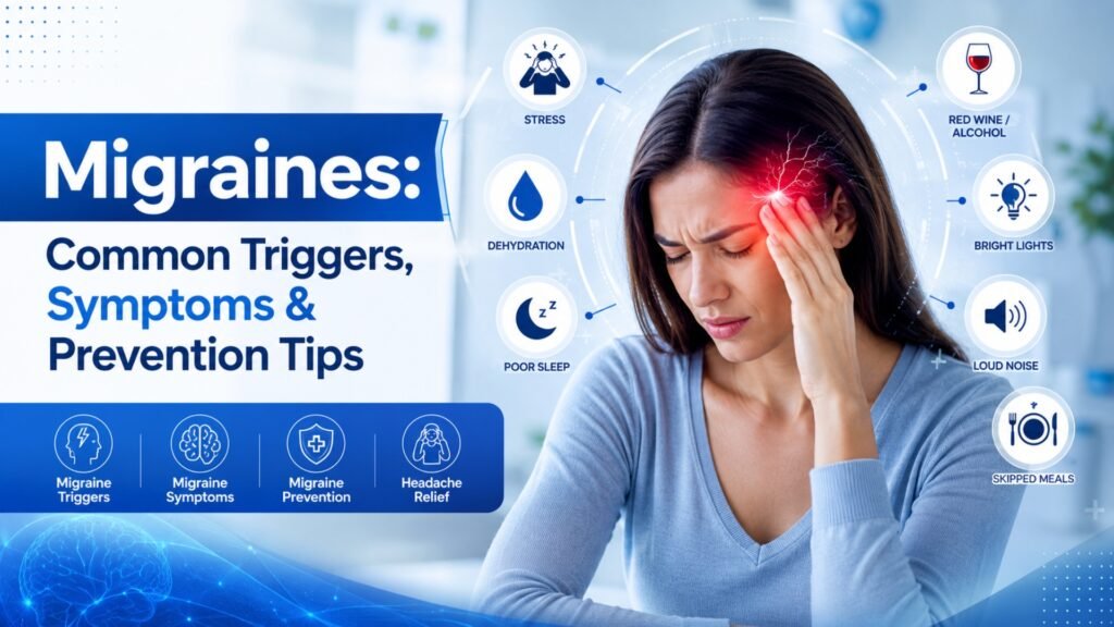
Migraines: Common Triggers, Symptoms and Prevention Tips Migraine triggers are different for everyone, but almost every person who lives with migraines eventually learns that certain foods, habits, or environmental changes seem to set off an attack. If you have ever wondered why a stressful week, a skipped meal, or a glass of red wine left you in bed with a throbbing headache, you are not imagining it. Migraines are a real neurological condition, and understanding your personal migraine triggers is one of the most powerful steps you can take toward fewer, milder attacks. In the United States, migraines affect people of nearly every age group, though they are most common among adults between 18 and 44, and women are affected roughly three times more often than men. Severity tends to vary based on overall health, hormonal status, stress levels, and underlying conditions such as high blood pressure or sleep disorders. This guide walks through what migraines are, the most common migraine triggers, the symptoms that signal an attack is coming, and prevention strategies that actually work. What Is a Migraine, Exactly? A migraine is not “just a bad headache.” It is a neurological event that typically causes moderate to severe, pulsating pain, often on one side of the head. Migraines frequently come with nausea, vomiting, and heightened sensitivity to light and sound. Some people also experience an “aura” — visual disturbances like flashing lights or blind spots — before the pain even begins. Migraines can last anywhere from a few hours to several days, and for many people they are disabling enough to disrupt work, school, and family life. If headaches are interfering with your quality of life, it is worth getting a proper evaluation. You can read more about how different headache types are diagnosed in our related post, Tension Headaches vs. Migraines: How to Tell the Difference. Migraines: Who Is Most Affected? Migraine prevalence and severity shift across the lifespan: Children and teens: Migraines can begin as early as childhood, though they often present differently, with shorter attacks and more stomach-related symptoms. Adults 18–44: This is the peak age range for migraine frequency, often tied to work stress, hormonal cycles, and lifestyle factors. Women during reproductive years: Hormonal fluctuations make this group especially prone to migraines, particularly around menstruation. Adults over 50: Migraine frequency often declines with age, but severity and the risk of related health complications, like cardiovascular issues, can increase. Underlying health conditions also play a major role in severity. People managing high blood pressure, chronic stress, sleep apnea, or hormonal imbalances tend to experience more frequent and more intense migraine attacks. Common Migraine Triggers Identifying your personal migraine triggers is one of the most effective tools for prevention. While triggers vary from person to person, research and clinical experience point to several categories that affect most migraine sufferers. Dietary Migraine Triggers Aged cheeses and processed meats containing tyramine Alcohol, especially red wine Caffeine — both excessive intake and sudden withdrawal Artificial sweeteners and MSG Skipped meals or prolonged fasting Environmental and Sensory Triggers Bright or flickering lights Strong smells, including perfume, smoke, or cleaning products Sudden weather changes or barometric pressure shifts Loud noises or crowded environments Lifestyle and Physical Triggers Chronic stress or sudden emotional shifts (even relief after stress can trigger an attack) Poor sleep quality or irregular sleep schedules Dehydration Intense physical exertion Hormonal changes, particularly drops in estrogen during the menstrual cycle Why Tracking Triggers Matters Because migraine triggers are highly individual, keeping a simple headache diary — noting food, sleep, stress levels, and weather — can reveal patterns that are easy to miss otherwise. Many patients are surprised to discover that their attacks cluster around a specific combination of factors rather than a single cause. Recognizing Migraine Symptoms Early Migraines often unfold in four distinct phases, and recognizing early symptoms can help you intervene before the pain becomes severe. Phase Typical Symptoms Duration Prodrome Mood changes, food cravings, neck stiffness, fatigue Hours to 1–2 days before Aura (in some people) Visual flashes, blind spots, tingling sensations 5–60 minutes Attack Throbbing pain, nausea, light/sound sensitivity 4–72 hours Postdrome Exhaustion, difficulty concentrating, mild residual pain Up to 24 hours after Not everyone experiences all four phases, and aura occurs in only about a quarter of migraine cases. Still, learning to recognize your own prodrome symptoms can give you a valuable window to act — whether that means taking medication, resting in a dark room, or simply removing yourself from a triggering environment. If your headaches are accompanied by confusion, vision loss, severe sudden onset (“the worst headache of your life”), fever, neck stiffness, or symptoms following a head injury, seek emergency medical care immediately. These can indicate a more serious underlying issue that needs urgent attention. Migraine Prevention Tips That Actually Work Prevention is rarely about a single fix — it is about consistency across several areas of daily life. Stick to a regular sleep schedule. Going to bed and waking up at the same time every day, including weekends, helps stabilize the nervous system. Stay hydrated. Dehydration is one of the most overlooked migraine triggers, and something as simple as drinking water consistently throughout the day can reduce frequency. Eat at regular intervals. Skipping meals causes blood sugar swings that can provoke an attack. Limit known dietary triggers. Once you identify your personal food triggers through a headache diary, reducing or eliminating them can make a noticeable difference. Manage stress proactively. Techniques such as deep breathing, gentle exercise, and mindfulness can lower the frequency of stress-related migraines. Moderate caffeine and alcohol intake. Consistency matters more than total elimination for many people. Consider preventive medication or supplements. For people with frequent or severe migraines, a healthcare provider may recommend daily preventive treatment, magnesium, riboflavin, or other evidence-based options. Protect your eyes from screen strain. Frequent breaks and blue-light filtering can reduce visually triggered migraines, especially for people who spend long hours at a computer. For more on this, see our post
Ischemic Stroke: Early Warning Signs, Treatment and Recovery
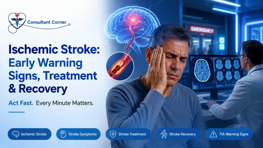
Ischemic Stroke: Early Warning Signs, Treatment and Recovery Ischemic stroke is one of the most time-sensitive medical emergencies a person can face, and recognizing it within seconds can be the difference between full recovery and permanent disability. An ischemic stroke occurs when a blood vessel supplying the brain becomes blocked by a clot, cutting off oxygen and nutrients to brain tissue. Within minutes, brain cells begin to die, which is why doctors often say “time is brain.” Understanding the early warning signs, available treatments, and what recovery actually looks like can help you or a loved one act fast and get the right care when it matters most. In the United States, stroke remains one of the leading causes of death and long-term disability, and ischemic stroke accounts for the vast majority of all stroke cases. This disease is more common in certain age groups — particularly adults over 55 — but it can strike younger people too, especially those with uncontrolled blood pressure, diabetes, or heart conditions. Its severity often depends heavily on the underlying health of the individual, how quickly symptoms are recognized, and how fast appropriate treatment begins. This guide walks through everything you need to know about ischemic stroke: how to spot it early, why immediate evaluation saves lives, who is most at risk, what treatment looks like, and how recovery unfolds. If you’re looking for related reading, you may also want to check out our posts on Understanding TIA: The Warning Stroke You Shouldn’t Ignore and Managing High Blood Pressure to Protect Your Brain. What Is an Ischemic Stroke? An ischemic stroke happens when a blood clot or fatty deposit blocks an artery that carries blood to the brain. Roughly 87% of all strokes are ischemic, according to the American Stroke Association. Unlike a hemorrhagic stroke, which involves bleeding in the brain, an ischemic stroke is caused by an obstruction that starves brain tissue of oxygen-rich blood. The longer that blockage persists, the more brain cells die — which is why rapid diagnosis and treatment are critical. There are two primary types of ischemic stroke: Thrombotic stroke — a clot forms directly inside an artery in the brain, often where the artery has narrowed due to plaque buildup (atherosclerosis). Embolic stroke — a clot forms elsewhere in the body (often the heart) and travels through the bloodstream until it lodges in a brain artery. Both types produce similar symptoms and require emergency evaluation. Why Ischemic Stroke Is a True Medical Emergency Every minute an ischemic stroke goes untreated, the brain loses an estimated 1.9 million neurons. That’s why emergency departments and stroke centers are built around speed — every test, every decision, and every treatment window is measured in minutes, not hours. Recognize the Warning Signs Early Ischemic strokes strike suddenly, often without warning. If you notice any of these symptoms — even if they come and go — get emergency help immediately: Sudden weakness or numbness on one side of the face, arm, or leg Trouble speaking or understanding words Sudden vision loss or blurriness Dizziness, imbalance, or sudden trouble walking Intense, sudden headache with no known cause Use the FAST Test The FAST acronym is the simplest way to remember the warning signs of stroke: F — Face drooping A — Arm weakness S — Speech difficulty T — Time to call 911 These symptoms are your body’s alarm system. They are not something to “wait out” or sleep off — every ischemic stroke symptom deserves an immediate call to emergency services. Why Immediate Stroke Evaluation Saves Lives Acting quickly after stroke symptoms appear can determine whether a patient is eligible for time-sensitive treatments, including: Clot-busting medication (tPA), which must typically be given within 3 to 4.5 hours of symptom onset Mechanical clot removal (thrombectomy), which can be effective for certain large-vessel blockages within a longer window Advanced stroke prevention therapy to reduce the risk of a second event Even brief, fleeting symptoms can represent a TIA (transient ischemic attack), sometimes called a “mini-stroke.” A TIA is often the final warning sign before a major ischemic stroke, and it should never be ignored simply because symptoms resolved on their own. A fast evaluation after a TIA can prevent a far more serious, disabling stroke down the line. Treatment Window Intervention Best Candidate 0–4.5 hours from onset tPA (clot-busting medication) Confirmed ischemic stroke, no bleeding risk Up to 24 hours (select cases) Mechanical thrombectomy Large-vessel blockage confirmed on imaging Ongoing Blood pressure & cholesterol management All ischemic stroke and TIA patients Ongoing Antiplatelet or anticoagulant therapy Patients with clotting risk or atrial fibrillation Long-term Rehabilitation (physical, speech, occupational) Patients with post-stroke deficits Are You at Risk for an Ischemic Stroke? Some risk factors for ischemic stroke can be managed, while others cannot. Knowing your personal risk profile is one of the most powerful tools for prevention. Common risk factors include: High blood pressure (the No. 1 cause of stroke) Diabetes High cholesterol Atrial fibrillation or other heart disease Smoking or vaping Obesity and a sedentary lifestyle Previous stroke or TIA Family history of stroke According to the Centers for Disease Control and Prevention, nearly 80% of strokes are preventable through management of these risk factors. If you have one or more of these conditions, scheduling an early neurology consultation truly matters — prevention is almost always more effective, and less costly, than treating a stroke after it happens. You might also find it helpful to read our related article on How Atrial Fibrillation Increases Stroke Risk for a deeper look at the heart-brain connection. How Ischemic Stroke Is Diagnosed When someone arrives at the hospital with suspected stroke symptoms, doctors move quickly through a standardized evaluation process. This typically includes: A rapid neurological exam to assess speech, vision, strength, and coordination CT or MRI imaging of the brain to confirm whether the stroke is ischemic or hemorrhagic Blood tests to check clotting factors, glucose, and other markers Imaging of the blood vessels (CT
Head Injury Explained: Symptoms, Recovery and Medical Care
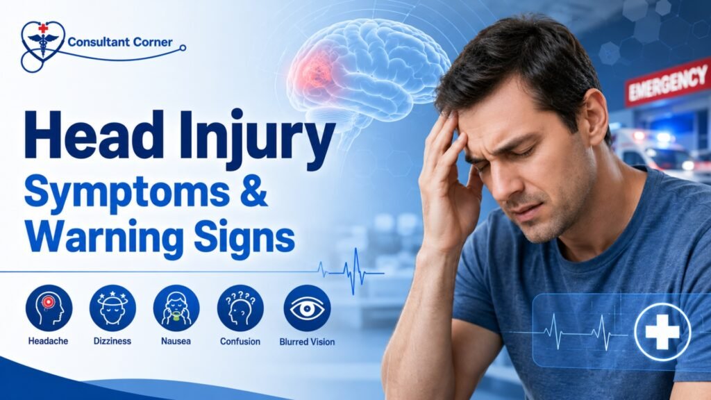
Head Injury Explained: Symptoms, Recovery and Medical Care A head injury can happen in the blink of an eye — a bathroom fall, a collision on the road, a sports impact, or even an accidental knock against a cabinet door. But the effects can ripple outward through days, weeks, or even months of your life. Understanding what a head injury actually means, knowing which symptoms signal danger, learning what recovery really looks like, and recognizing when to call a doctor can make a profound difference in how fully — and how quickly — you or someone you love recovers. This comprehensive guide covers everything you need to know about head injury: the different types, the common and emergency warning signs, the full stages of recovery, the special populations most at risk, how doctors make a diagnosis, and the practical steps you can take right now to protect your brain health. What Is a Head Injury? A head injury is any form of trauma to the scalp, skull, or brain. The spectrum is wide — ranging from a minor bump that leaves a small lump and fades within a day, all the way to a traumatic brain injury (TBI) that disrupts memory, mood, movement, or consciousness for months or longer. Not every head injury directly involves the brain. A scalp laceration may bleed dramatically because scalp tissue is highly vascular, yet involve zero brain trauma. Conversely, a head injury that appears minor on the surface can still cause significant internal damage to the brain beneath the skull. Medically, head injuries fall into two primary categories: Closed Head Injuries The skull remains intact, but the brain can still be jolted, bruised, or twisted inside the cranial cavity. The most common type is a concussion, which occurs when the brain moves rapidly within the skull and temporarily disrupts normal brain function. Closed head injuries account for roughly 75% of all traumatic brain injuries in the United States each year. Open (Penetrating) Head Injuries An object breaches the skull and may directly damage brain tissue. These are generally more severe, almost always require emergency surgical intervention, and carry a significantly higher risk of permanent neurological damage. Within both categories, a head injury is further classified by severity — mild, moderate, or severe — based on whether the person lost consciousness, how long confusion lasted, and what brain imaging reveals. How Common Is Head Injury? Head injury is far more common than most people realize. According to the Centers for Disease Control and Prevention (CDC), approximately 1.7 million traumatic brain injuries occur in the United States each year, with about 3% resulting in death. Adults are the most frequently affected group, though children and older adults face their own heightened risks that deserve special attention. Common Causes of Head Injury The cause of a head injury often shapes the type and severity of the damage that follows. Understanding the mechanism matters, because high-energy impacts — such as those in motor vehicle accidents or falls from height — carry a much higher likelihood of serious brain injury even when initial symptoms appear mild. The most common causes include: Falls — Falls are the single leading cause of head injury overall, particularly in young children and adults over age 65. Poor balance, vision changes, and medications that impair coordination all significantly elevate fall risk in older adults. Motor Vehicle Accidents — The forces involved in car collisions tend to produce more severe injuries. Whiplash-type movements can cause the brain to strike the interior of the skull even when there is no direct head impact. Sports-Related Impacts — Contact sports like football, soccer, rugby, hockey, and boxing frequently produce concussions. Even cycling and skiing carry significant head injury risk without a properly fitted helmet. Workplace Accidents — Construction and industrial workers face elevated risk from falling objects, falls from elevated platforms, or machinery-related impacts. Physical Assaults — Blunt force trauma to the head during an assault accounts for a meaningful percentage of emergency department head injury visits each year. Head Injury Symptoms: What to Watch For One of the most critical things to understand about a head injury is that symptoms do not always appear immediately. They can emerge hours — sometimes even a day or two — after the initial trauma. This delayed onset is precisely why anyone who sustains a head injury should be monitored closely for at least 24 to 48 hours, even when they feel completely fine right after the incident. Common Symptoms of a Head Injury Persistent or worsening headache Dizziness or problems with balance and coordination Nausea or vomiting Confusion, mental fog, or difficulty concentrating Memory problems, especially difficulty recalling the injury event itself (post-traumatic amnesia) Sensitivity to light (photophobia) or noise (phonophobia) Unusual fatigue or drowsiness Irritability, mood swings, or heightened anxiety Ringing in the ears (tinnitus) or blurred vision Disrupted sleep — either difficulty falling asleep or sleeping far more than usual These symptoms overlap considerably with other neurological conditions. For instance, a persistent headache after a head injury can look very similar to a migraine disorder. If you’ve been experiencing recurrent headaches, our related guide — Can Bad Posture Cause Headaches? Causes, Symptoms & Treatment — explores how different structural triggers can produce overlapping symptoms. Emergency Warning Signs After a Head Injury While many head injury symptoms can be managed with rest and careful at-home monitoring, certain signs demand immediate emergency care. Brain swelling and internal bleeding can develop without any visible external injury, and these are conditions that can become life-threatening very rapidly. Call 911 or Go to the Emergency Room Immediately If: The headache rapidly intensifies rather than easing There is repeated or forceful vomiting (more than once or twice) The person has slurred speech or sudden difficulty finding words A seizure or convulsion occurs One pupil is noticeably larger than the other (unequal pupils) The person shows growing confusion, extreme disorientation, or dramatic behavioral change There is any loss of consciousness, even briefly One
Post-Stroke Recovery & Rehabilitation
How the Brain Heals and What Truly Drives Recovery A Neurologist-Guided, Patient-Centered Roadmap Stroke Recovery Is a Neurological Process — Not a Waiting Period A stroke is not simply a sudden medical event; it is a neurological injury that disrupts the brain’s ability to control movement, speech, cognition, behavior, and vital bodily functions. Whether caused by an ischemic blockage or an intracerebral hemorrhage, stroke results in acute damage to brain tissue followed by a complex and evolving recovery process. In the United States, nearly 800,000 people experience a stroke each year, making it one of the leading causes of long-term neurological disability. Advances in acute stroke treatment have significantly improved survival. However, survival is only the beginning. What follows is a prolonged and highly individualized process of neurological recovery. For patients and families, this phase is often filled with uncertainty:Will strength return? Will speech improve? How long does recovery take? Is rehabilitation truly effective? From a neurological standpoint, recovery is best understood through two core principles: Stroke recovery is driven by brain plasticity, not time alone Rehabilitation is an active neurological treatment, not supportive care Understanding these principles helps patients and caregivers move forward with clarity, realistic expectations, and hope. How the Brain Heals After Stroke: Understanding Neuroplasticity When a stroke occurs, part of the brain is injured or deprived of blood flow, leading to loss of neurons and disruption of neural networks. Importantly, this injury does not mean the brain is incapable of recovery. Instead, recovery depends on how surviving brain regions adapt over time — a process known as neuroplasticity. What Is Neuroplasticity? Neuroplasticity refers to the brain’s ability to reorganize, strengthen existing connections, and form new neural pathways in response to injury and experience. After a stroke, unaffected areas of the brain can partially assume functions previously handled by damaged regions. This biological adaptability is the foundation of rehabilitation. Crucially, neuroplasticity is activity-dependent. The brain does not reorganize simply with rest or time. Recovery is driven by repetitive, meaningful, task-specific practice, which is why structured rehabilitation is so effective. Early and Late Brain Recovery In the first days to weeks after a stroke, the brain enters a period of heightened plasticity. Swelling subsides, inflammation decreases, and neural signaling stabilizes. During this phase, early improvements may reflect recovery of stunned but viable tissue. While the most rapid gains often occur within the first 3–6 months, recovery does not stop there. Research shows that meaningful improvement can continue for 6–12 months and beyond, particularly when rehabilitation remains progressive and goal-directed. Later recovery often involves: Refinement of motor control rather than large strength gains Improved balance, coordination, and endurance Gradual improvements in language and cognition Development of compensatory strategies that enhance independence The idea that recovery “plateaus” early is often a reflection of reduced therapy intensity or lack of reassessment — not a true limit of neurological potential. Rehabilitation Therapies That Drive Recovery Stroke rehabilitation is not a single therapy or a one-size-fits-all program. It is a multidisciplinary neurological intervention designed to retrain the brain by engaging impaired neural networks repeatedly and purposefully. Physical Therapy: Restoring Mobility and Balance Physical therapy focuses on improving strength, coordination, balance, and gait. Weakness, spasticity, and impaired balance are common after a stroke and significantly affect independence. Through task-oriented and repetitive movement training, physical therapy helps the brain re-map motor pathways, reduce fall risk, and improve functional mobility. Therapy evolves — from basic transfers to higher-level balance, endurance, and community mobility. Occupational Therapy: Regaining Independence in Daily Life Occupational therapy helps patients regain the ability to perform activities of daily living, such as dressing, bathing, eating, writing, and managing household tasks. Stroke often affects fine motor control, visual-spatial processing, and executive function — all critical for daily independence. OT integrates motor, sensory, and cognitive systems, reinforcing purposeful use of affected limbs and promoting functional recovery rather than learned non-use. Speech and Language Therapy: Communication and Swallowing Stroke can affect language (aphasia), speech clarity (dysarthria), cognition, and swallowing (dysphagia). Speech-language therapy addresses: Language comprehension and expression Speech articulation and voice control Cognitive-communication skills Swallowing safety and nutrition Recovery in this domain may be gradual, but meaningful gains can continue long after motor recovery stabilizes. Early swallowing evaluation is essential to reduce aspiration risk and medical complications. Cognitive Rehabilitation: Attention, Memory, and Executive Function Cognitive changes after stroke are common and frequently underrecognized. Patients may struggle with attention, memory, processing speed, or executive skills — even when physical recovery appears strong. Cognitive rehabilitation helps patients develop new strategies to strengthen preserved neural pathways and compensate for deficits. Recovery in this domain often continues later into the recovery process, reinforcing the importance of ongoing reassessment. Early Recovery: The First Weeks Matter — But Don’t Define Everything The first days to weeks after a stroke represent a critical window of opportunity. During this phase, early mobilization and rehabilitation — when medically appropriate — improve functional outcomes and reduce complications. That said, early appearance does not define long-term outcome. Neurologists caution against premature prognostication. Patients may appear severely impaired early on yet make meaningful gains over time, while others with milder strokes may struggle due to secondary complications. Common early challenges include: Fatigue Fluctuating weakness or tone Speech or swallowing difficulty Cognitive slowing or confusion Emotional distress These challenges are common and treatable. Addressing them early improves participation in rehabilitation and long-term outcomes. What Really Predicts Stroke Recovery Recovery is shaped by multiple interacting factors — not a single variable. What Matters Most: Quality, timing, and intensity of rehabilitation Patient engagement and education Ongoing neurological oversight Social and caregiver support Management of medical and neurological complications What Matters Less Than Commonly Believed: Age alone (older adults retain neuroplastic capacity) Early severity as a fixed predictor Arbitrary recovery timelines From a neurologist’s perspective, trajectory over time is far more meaningful than a single snapshot. Cognitive, Emotional, and Behavioral Recovery Physical recovery is often the most visible aspect of stroke rehabilitation, but cognitive and emotional changes can be equally impactful. Cognitive Changes Difficulties with attention, memory, executive function, or visual-spatial processing may affect independence, work, and social engagement. These deficits may emerge
7 Subtle Stroke Signs in Women You Might Be Missing
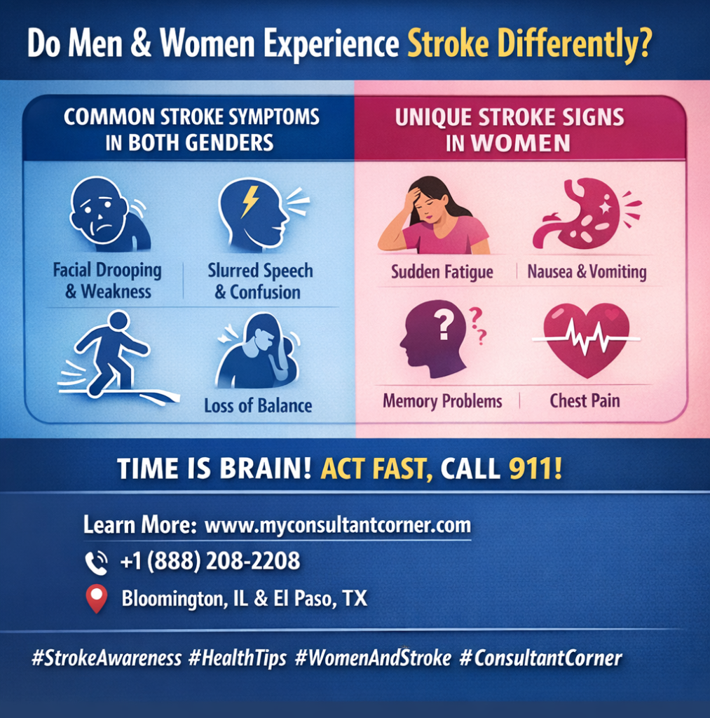
Do Men and Women Experience Stroke Differently? While the core mechanics of a stroke remain the same, clinical data show significant differences in how symptoms manifest based on gender. According to the World Stroke Organization (WSO), women are not only more likely to experience a stroke in their lifetime. Still, they are also 11% more likely to report non-traditional symptoms that lead to delayed diagnosis. Understanding these nuances is vital because every 60 seconds of delayed treatment results in the death of nearly 2 million brain cells. What Common Symptoms Are Shared by Both Genders? In the majority of cases, both men and women will display the “classic” neurological deficits associated with brain cell death. These symptoms are primarily focused on motor skills and sensory processing: Sudden Asymmetry: Drooping of the face, or paralysis of the arm or leg—typically isolated to one side of the body. Cognitive & Sensory Disruptions: Slurred speech, sudden confusion, vision loss, or a severe headache with no identifiable cause. Mobility Issues: Difficulty walking or a sudden loss of balance (the “B” in the BE FAST protocol). Why Are Stroke Signs Frequently Missed in Women? Women are more likely to present with “subtle” or nonspecific symptoms, which are often mistakenly attributed to stress, exhaustion, or other minor ailments. This diagnostic gap is a leading cause of poorer outcomes for female patients. Common “Non-Traditional” Symptoms in Women: Systemic Weakness: A feeling of generalized exhaustion or extreme fatigue that comes on suddenly. Gastrointestinal & Respiratory Signs: Nausea, vomiting, hiccups, or shortness of breath. Altered Mental Status: Disorientation, sudden memory problems, or acting “out of character”. Cardiac Mimics: Chest pain, palpitations, or a racing heartbeat that can sometimes be confused with a heart attack. Which Symptoms Are Relatively More Common in Men? Research suggests men are more likely to exhibit specific coordination and sensory issues. While these are still “classic” signs, they appear with higher frequency in male patients: Ataxia: A sudden, noticeable loss of coordination in the limbs or an unsteady gait. Paresthesia: Distinct sensations of numbness, tingling, or “pins and needles”. Visual Disturbances: Higher reported rates of double vision or sudden dimming of sight in one eye. How Does Gender Affect Treatment Timing and Survival? Feature Statistical Impact on Women Impact on Recovery Recognition Nonspecific signs (nausea, fatigue) lead to frequent misdiagnosis. Delayed treatment. Response Time Women wait an average of 30 to 60 minutes longer to call 911. Increased brain damage. Age of Onset Women typically have strokes at an older age than men. Higher risk of mortality. Disability Risk Women face a higher rate of long-term nursing home placement post-stroke. Reduced independence. When Should You Seek Emergency Help? Regardless of sex, any sudden combination of these symptoms—even if they seem mild or resolve after a few minutes—must be treated as a medical emergency. Call Emergency Services (911): Do not wait for a clinic appointment or try to “sleep it off”. Trust Your Instincts: If a woman reports feeling “not right” combined with sudden fatigue or confusion, seek help immediately. Time is Brain: Accessing clot-busting therapies within the first 3–4.5 hours is the most effective way to prevent permanent disability. Your Recovery Doesn’t End at the Hospital Doors At Consultant Corner, we believe that surviving a stroke is only the beginning. We treat stroke recovery as a continuous journey, not a one-time medical event. Our specialized neurology team offers personalized, data-driven post-stroke care through both convenient virtual consultations and in-person clinic visits. We focus on neuroplasticity, secondary prevention, and restoring your independence. Take the Next Step Toward Healing If you or a loved one needs expert guidance on the path to recovery, our team is ready to support you. Visit: www.myconsultantcorner.com Call: +1 (888) 208-2208 Email: info@myconsultantcorner.com Address: 1404 Eastland Dr, Bloomington, IL 61701, United States Address: 5140 Montana Ave, El Paso, TX 79903, United States
5 Early Stroke Symptoms You Can’t Ignore (2026 Update)

What Are the Main Warning Signs of a Stroke? A stroke happens when blood flow to a part of the brain is interrupted, leading to brain cell damage within minutes. Recognizing the early warning signs can save your life or someone else’s. According to the World Health Organization (WHO), stroke is the second leading cause of death globally, responsible for over 12 million cases every year. Time is critical—knowing the signs and acting fast can reduce the risk of long-term disability and improve recovery outcomes. How Can You Identify Stroke Symptoms Quickly? (Remember: BE FAST) Medical experts use the BE FAST acronym to help people quickly recognize stroke symptoms. Each letter stands for a potential warning sign: B – Balance: Is There Sudden Dizziness or Loss of Coordination? If someone suddenly can’t walk straight, feels dizzy, or loses coordination, it could be an early stroke sign. Research from the American Stroke Association (ASA) suggests that around 40% of stroke patients experience balance problems or sudden dizziness. E – Eyes: Is There Trouble Seeing Clearly? Watch for blurred vision, double vision, or complete vision loss in one or both eyes. About 20% of stroke victims report sudden vision changes, which may appear alongside light sensitivity or visual field loss. F – Face: Is One Side Drooping? Ask the person to smile. If one side droops or feels numb, it’s a red flag. Facial asymmetry is one of the most common early signs of stroke and appears in over 60% of cases according to clinical data. A – Arm: Do the Arms Feel Weak or Numb? Have them raise both arms. If one arm drifts downward or can’t be lifted, that’s a strong indicator of a stroke. This weakness may also affect one leg, typically on the same side. S – Speech: Is the Speech Slurred or Confusing? Speech problems—like slurred words, difficulty understanding, or trouble finding the right words—often signal a stroke. Studies find that nearly 70% of stroke patients present some form of speech impairment at onset. T – Time: Why Is Every Minute Critical? If any of these signs appear, call emergency services immediately. Never wait for the symptoms to improve on their own. Each passing minute can cause an estimated 1.9 million brain cells to die, increasing the risk of permanent brain injury. What Are Other Sudden Symptoms Not to Ignore? Not every stroke follows the same pattern. Some people experience less common, but equally serious symptoms such as: Sudden, severe headache without a known cause—especially if it comes out of nowhere. Loss of consciousness, fainting, or seizure. Nausea, vomiting, or vertigo (a spinning sensation). Sudden confusion or trouble understanding speech. Always treat these as potential emergencies—even if symptoms fade after a few minutes. Why Is Early Recognition of Stroke So Important? When it comes to stroke, every minute truly counts. Each second of delay means more brain cells are lost, and long-term complications become more likely. The World Health Organization (WHO) reports that strokes cause over 6.5 million deaths globally each year, and the risk of severe disability rises sharply with every lost minute of treatment time. In short, the faster a stroke is recognized and treated, the better the outcome. Why Does Fast Action Improve Access to Treatments? Stroke treatments are highly time-sensitive. Clot-busting drugs (known as thrombolytics, such as tPA) and mechanical clot removal procedures must be given within 3 to 6 hours from symptom onset to be most effective. When strokes are recognized quickly: Patients reach CT scans faster, helping doctors confirm the stroke type (ischemic or hemorrhagic). Life-saving therapies can be started sooner, limiting brain damage. Supportive care (like managing blood pressure, oxygen, and glucose) can prevent secondary complications such as brain swelling or infection. In contrast, delayed recognition may mean missing the treatment window entirely, reducing the chance of recovery. How Does Early Recognition Improve Survival and Recovery? Evidence is clear: faster diagnosis leads to better survival and long-term independence. A large-scale study in The Lancet Neurology found that patients treated within the first 90 minutes of a stroke were three times more likely to recover without major disability. Prompt recognition and treatment also mean: Lower mortality rates. Fewer hospital complications, such as blood clots and pneumonia. Greater chances of walking and speaking normally after recovery. Essentially, time to treatment predicts quality of life after stroke. When Should You Go to the ER for a Stroke? If you suspect a stroke—even slightly—call emergency services immediately. Don’t wait to see if symptoms improve, and don’t try to drive yourself. Stroke is a time‑critical medical emergency, and fast action can mean the difference between full recovery and permanent disability. According to the American Heart Association, early treatment within the first few hours can reduce disability by up to 30% and significantly improve survival. The Consultant Corner Approach to Ischemic Stroke At Consultant Corner, we view ischemic stroke as an ongoing recovery process, not a single event. Our neurology team provides personalized post-stroke care through both in-clinic and virtual visits, focusing on recovery, prevention, and long-term brain health. Take the Next Step If you or a loved one is recovering from an ischemic stroke, we’re here to help.Visit www.myconsultantcorner.com, call: +1 (888) 208-2208, or Email: info@myconsultantcorner.com to get started. Address: 1404 Eastland Dr, Bloomington, IL 61701, United States Address: 5140 Montana Ave, El Paso, TX 79903, United States
Syncope & Fainting Spells: Neurological vs Non-Neurological Causes and Expert Care
Syncope & Fainting Spells Neurological vs Non-Neurological Causes, Diagnostic Evaluation, and Expert Care A Comprehensive, Patient-Centered Guide from Consultant Corner Passing out—even briefly—is one of the most unsettling experiences a person can have. Patients often describe fainting spells as sudden, unpredictable, and frightening, while families immediately worry about what it might mean: Was it a seizure? A heart rhythm problem? A stroke? That uncertainty is understandable, because “passing out” can represent everything from a benign reflex episode to a potentially life-threatening cardiac condition—or a neurological event that was mistaken for fainting. Clinically, syncope is common and frequently prompts urgent medical evaluation. It accounts for approximately 1–3% of emergency department visits and up to 6% of hospital admissions, highlighting both its prevalence and the challenge of safely identifying high-risk patients. Over a lifetime, at least one-third of individuals will experience syncope, with incidence rising again in older adulthood—precisely the population at greatest risk for injury, medication interactions, autonomic dysfunction, and cardiac disease. In many cases, patients are reassured quickly and discharged without a clear explanation. Sometimes that reassurance is appropriate. In other cases, however, premature diagnostic closure can miss a serious underlying condition or delay appropriate follow-up. The central challenge is that syncope is not a diagnosis—it is a symptom. It reflects a brief, reversible reduction in blood flow to the brain, and the clinician’s task is to determine why that reduction occurred and whether the patient is at risk of recurrence, injury, or sudden deterioration. From a neurologist’s perspective, fainting spells sit at the crossroads of neurology, cardiology, and autonomic physiology. Many events described as “fainting” are not true syncope at all, but rather neurological mimics such as seizures or transient alterations in awareness. At the same time, some of the most dangerous causes are cardiac and may present with little or no warning. The goal of evaluation is to separate neurological from non-neurological causes, identify red flags early, and pursue targeted—not excessive—testing. At Consultant Corner, we approach syncope as a diagnostic problem that deserves clarity, evidence-based reasoning, and individualized care. Whether an episode represents vasovagal syncope, orthostatic hypotension, arrhythmia, seizure, or autonomic dysfunction, our focus is on helping patients understand what happened, what risks matter, and how to move forward safely. What Is Syncope — and Why the Brain Is Central Syncope is defined as a transient loss of consciousness due to temporary, global cerebral hypoperfusion, followed by rapid and complete recovery. In practical terms, fainting occurs when the brain is briefly deprived of sufficient oxygen and glucose to maintain consciousness. Unlike coma or traumatic brain injury, syncope does not involve permanent brain damage; however, it signals a failure in the systems that normally sustain cerebral perfusion. The brain is uniquely sensitive to even small changes in blood pressure and cardiac output. Consciousness depends on tightly regulated interactions between the heart, blood vessels, and autonomic nervous system. When these systems fail—due to sudden hypotension, abnormal heart rhythm, impaired autonomic reflexes, or volume depletion—cerebral perfusion can fall below a critical threshold within seconds, resulting in collapse. What makes syncope challenging is that it represents a final common pathway rather than a single disease. Unlike seizures or strokes, syncope does not originate from intrinsic electrical or structural brain pathology. Instead, it reflects failure of the physiological support systems that maintain brain function. In clinical practice, however, this distinction is often blurred, as neurological disorders may mimic syncope, trigger syncope, or coexist with non-neurological causes. Neurological vs Non-Neurological Causes: Why the Distinction Matters One of the most important steps in evaluating fainting spells is determining whether the underlying cause is neurological or non-neurological. This distinction directly shapes diagnostic testing, treatment decisions, long-term risk assessment, and patient counseling. From a neurologist’s standpoint, the priority is not merely confirming that syncope occurred, but ensuring that neurological conditions are not overlooked, particularly those that masquerade as syncope or present with transient loss of consciousness as a secondary manifestation. At the same time, it is equally important to recognize when fainting is driven by cardiac or systemic disease that carries its own risks. Because symptoms often overlap and witness descriptions are frequently incomplete, fainting spells require a structured, neurologist-led approach that integrates neurological, cardiovascular, and autonomic factors rather than evaluating them in isolation. Neurological Causes and Neurologic Mimics of Syncope Seizures: The Most Clinically Significant Mimic Distinguishing syncope from epileptic seizures is one of the most consequential challenges in evaluation. Both can present with sudden collapse and loss of consciousness, but the safety implications, driving restrictions, employment, and long-term management differ substantially. Seizures arise from abnormal, hypersynchronous electrical activity in the brain and are typically followed by a post-ictal period characterized by confusion, fatigue, headache, or focal neurological deficits. Syncope, in contrast, results from transient cerebral hypoperfusion and is usually followed by rapid recovery once blood flow is restored. In practice, overlap is common. Syncope may be accompanied by brief involuntary movements due to brainstem hypoxia (convulsive syncope), while some seizures may be brief or subtle and lack dramatic convulsions. Because missing a seizure diagnosis carries serious consequences, careful neurological assessment is essential, and EEG or prolonged monitoring may be required when history alone is insufficient. Autonomic Nervous System Disorders and Neurogenic Syncope The autonomic nervous system continuously regulates blood pressure, heart rate, and vascular tone in response to posture, stress, and exertion. Disorders affecting this system can impair the body’s ability to maintain cerebral perfusion, leading to recurrent or unexplained syncope. Neurological conditions associated with autonomic failure include Parkinson’s disease and related neurodegenerative disorders, multiple system atrophy, diabetic autonomic neuropathy, small fiber neuropathies, and other disorders involving peripheral or central autonomic pathways. In recent years, post-viral and post-COVID dysautonomia have emerged as increasingly recognized causes of syncope and orthostatic intolerance, particularly in younger patients without structural heart disease. Neurogenic syncope is often posture-dependent, recurrent, and accompanied by other autonomic symptoms such as heat intolerance, gastrointestinal dysmotility, urinary dysfunction, or abnormal sweating. Diagnosis frequently requires specialized autonomic testing and longitudinal neurological care, rather than episodic reassurance. Structural and Cerebrovascular Neurological Considerations Although uncommon, certain structural or vascular neurological conditions can present with transient loss of consciousness.
Hand Numbness & Carpal Tunnel Syndrome | When to See a Neurologist

Carpal Tunnel Syndrome & Nerve Entrapment Disorders Understanding Symptoms, Diagnosis, and When Neurological Evaluation Matters A Patient-Centered, Evidence-Based Guide from Consultant Corner Numbness, tingling, pain, or weakness in the hands and arms are among the most common neurological complaints encountered in outpatient practice. Patients often describe their hands “falling asleep,” waking at night with burning or pins-and-needles sensations, dropping objects unexpectedly, or struggling with fine motor tasks such as typing, writing, or buttoning clothes. These symptoms can be disruptive, frightening, and persistent — and they often raise an important question: Is this something serious? Carpal tunnel syndrome is frequently the first diagnosis patients encounter when they search for answers online. It is indeed common, affecting an estimated 3–6% of adults, and it represents the most frequent peripheral nerve entrapment disorder. However, not all hand numbness is carpal tunnel syndrome, and not all nerve symptoms originate at the wrist. From a neurologist’s perspective, symptoms involving the hands and arms sit at the intersection of peripheral nerve anatomy, spinal health, systemic disease, occupational factors, and individual anatomy. This complexity explains why nerve entrapment disorders are sometimes dismissed too quickly — or, conversely, overdiagnosed and overtreated. Accurate diagnosis requires careful localization, clinical reasoning, and, in many cases, objective testing. At Consultant Corner, we approach carpal tunnel syndrome and related nerve entrapment disorders with a structured, evidence-based neurological framework designed to provide clarity, reassurance, and appropriate care. Understanding Nerve Entrapment: A Neurological Perspective Peripheral nerves travel long distances from the spinal cord to the muscles and skin, passing through narrow anatomical corridors formed by bones, ligaments, and soft tissue. At certain predictable locations, these nerves are vulnerable to compression. Even modest increases in pressure — due to inflammation, fluid retention, repetitive motion, or structural crowding — can disrupt nerve blood flow and electrical signaling. This process, known as nerve entrapment, typically develops gradually. Early symptoms are often sensory, such as intermittent tingling or numbness. If compression persists, symptoms may become constant and progress to weakness, coordination difficulty, or muscle atrophy. Importantly, symptoms are not always felt at the site of compression, which is why localization based on anatomy — rather than symptom location alone — is central to diagnosis. Carpal Tunnel Syndrome: Common, but Often Misunderstood Carpal tunnel syndrome results from compression of the median nerve as it passes through the carpal tunnel at the wrist. This tunnel is a rigid, confined space, and it has very little capacity to accommodate swelling or structural change. As pressure increases, the median nerve becomes vulnerable to dysfunction. Patients with carpal tunnel syndrome typically experience numbness, tingling, or burning sensations in the thumb, index finger, middle finger, and part of the ring finger. Symptoms often worsen at night or during activities involving sustained wrist positioning. Many patients report relief by shaking or repositioning the hand — a classic feature that reflects temporary restoration of nerve blood flow. As compression progresses, patients may develop weakness of thumb movements, difficulty with fine motor tasks, or frequent dropping of objects. These signs suggest more advanced nerve involvement and warrant timely evaluation. A key anatomical clue is that carpal tunnel symptoms usually spare the little finger, which is supplied by a different nerve. Carpal tunnel syndrome may exist on its own, but it can also coexist with cervical spine disease or generalized neuropathy — a phenomenon sometimes referred to as double crush syndrome. In these cases, symptoms may be more severe or less responsive to isolated wrist treatment, reinforcing the importance of a comprehensive neurological assessment. Other Nerve Entrapments That Mimic Carpal Tunnel Syndrome While carpal tunnel syndrome is the most common diagnosis, many patients referred for “carpal tunnel–like” symptoms ultimately have nerve compression at a different site, or at multiple sites. The ulnar nerve, for example, is frequently compressed at the elbow in cubital tunnel syndrome. This condition typically causes numbness in the ring and little fingers, hand weakness, and symptoms worsened by prolonged elbow flexion or pressure. Compression can also occur at the wrist in Guyon’s canal, producing a different but related symptom pattern. The radial nerve may be compressed in the forearm, leading to pain or weakness of wrist and finger extension, often without prominent sensory symptoms. In the lower extremities, compression of the peroneal nerve near the knee can cause foot drop, while tarsal tunnel syndrome involves compression of the posterior tibial nerve at the ankle, producing burning or tingling in the sole of the foot. These conditions illustrate why accurate localization matters. Treatments that help carpal tunnel syndrome may not improve symptoms caused by ulnar or radial nerve compression, and unnecessary procedures can be avoided with careful neurological evaluation. Neurological, Orthopedic, and Systemic Contributors: Why Symptoms Overlap Hand and limb symptoms do not always arise from focal nerve entrapment. Orthopedic conditions such as arthritis or tendon disease may cause pain and stiffness that limit function but do not follow nerve distributions. Systemic conditions — including diabetes, thyroid disease, vitamin deficiencies, autoimmune disorders, and medication effects — can produce diffuse nerve dysfunction that mimics entrapment. Neurologists differentiate these possibilities by focusing on pattern recognition: nerve-specific sensory changes, reflex alterations, weakness patterns, and progression over time. This distinction is critical because treating a single compression site will not resolve symptoms driven by a systemic process. When Symptoms Should — and Should Not — Raise Concern Many nerve entrapment symptoms are intermittent and reversible, especially early in their course. Tingling that occurs primarily at night, symptoms triggered by certain positions, or discomfort relieved by rest or splinting are often signs of functional nerve compression rather than permanent damage. However, persistent numbness, progressive weakness, visible muscle thinning, or loss of fine motor control warrant earlier neurological evaluation. Rapid progression, involvement of multiple limbs, or symptoms extending beyond a single nerve distribution raise concern for more proximal or systemic conditions. From a neurological standpoint, duration and progression matter more than intensity alone. Early evaluation allows for intervention before nerve injury becomes irreversible. How Nerve Entrapment Is Diagnosed A careful neurological examination remains the cornerstone of diagnosis. By assessing sensation, strength, reflexes, coordination, and muscle bulk, neurologists can often localize nerve
Epilepsy and Seizures: Diagnosis, Treatment, and Quality of Life

What Is Epilepsy and How Can It Change Your Life? Hey there, if you’ve ever wondered about epilepsy, you’re not alone. It’s one of the most common neurological conditions out there, affecting around 50 million people worldwide according to the World Health Organization. In the US alone, about 2.9 million adults live with active epilepsy—that’s roughly 1% of the adult population, per CDC data from 2021-2022. And get this: 1 in 26 people will develop epilepsy at some point in their life. But epilepsy isn’t just about dramatic seizures; it’s a complex brain disorder that touches everything from your daily routine to your long-term health. Let’s break it down in a way that’s easy to follow, like we’re chatting over coffee. Understanding Seizures vs. Epilepsy: What’s the Real Difference? First things first—seizures and epilepsy aren’t the same thing, and mixing them up can lead to confusion. A seizure is like a sudden electrical storm in your brain, often triggered by things like infections, injuries, or even alcohol withdrawal. But epilepsy? That’s when you have an ongoing tendency for unprovoked seizures. Research shows the annual incidence of epilepsy is about 61-68 cases per 100,000 people globally, based on meta-analyses from studies like those in the Neurology journal. If you’ve had just one seizure, it doesn’t automatically mean epilepsy. In fact, rushing to that label could mean unnecessary meds and stress. On the other hand, delaying a diagnosis may leave you vulnerable to additional risks. Think about it: accurate diagnosis helps predict recurrence—studies indicate that after a first unprovoked seizure, the risk of another can be as high as 40-50% without treatment. Why Is Epilepsy Considered a Brain Network Disorder? Epilepsy isn’t about one rogue neuron; it’s a whole network gone haywire. Your brain’s excitability increases, leading to those abnormal syncs. Causes vary—structural issues like strokes or tumors, genetic factors, inflammation, or even unknowns. Data from the Global Burden of Disease Study 2021 estimates 51.7 million people globally deal with this, with a prevalence of around 658 per 100,000. This network view explains why epilepsy looks different for everyone. Some have focal seizures starting in one brain area, while others are more widespread. Personalized care is key because, as research highlights, treating it like a one-size-fits-all just doesn’t cut it. How Do Epilepsy Seizures Really Show Up in Everyday Life? Forget the movie stereotypes—epilepsy seizures aren’t always full-body convulsions. Many are subtle: a brief stare, lip smacking, or that weird déjà vu feeling. Absence seizures in kids might just look like daydreaming, lasting seconds. These can mimic other issues like anxiety or migraines, leading to misdiagnosis. Witness accounts and your own story often tell more than tests. And stats back this: epilepsy affects all ages, but in older adults, new seizures often signal strokes or medication interactions. Early recognition matters because uncontrolled seizures disrupt work, school, and safety. Diagnosing Epilepsy: How Do We Get It Right Over Time? Diagnosing epilepsy is more art than science sometimes—it’s clinical, backed by tools like EEGs and MRIs. A normal EEG doesn’t rule it out, though; up to 50% of people with epilepsy have normal readings between seizures. Genetic tests or video monitoring help in tough cases. The process evolves with follow-ups. Longitudinal studies show that precise diagnosis boosts outcomes, reducing unnecessary treatments and improving life quality. Treating Epilepsy: Why Medication Adherence Is Your Best Friend For most with epilepsy, meds are the go-to. About 70% of people can achieve seizure freedom with proper treatment, per WHO findings. We pick based on your seizure type, age, and lifestyle—considering side effects and even pregnancy plans. But adherence? That’s huge. Missing doses causes breakthrough seizures, a top reason for ER visits. Research from longitudinal cohorts shows 50.5% get seizure-free on the first med, but consistency keeps it that way. Let’s talk openly about side effects; adjusting thoughtfully prevents bigger issues. How Do Anti-Seizure Meds Work in Managing Epilepsy? These meds tweak brain excitability—some block sodium channels to calm firing, others boost calming signals like GABA. Newer ones target multiple paths, expanding options for better control with fewer side effects. Data from 30-year studies: After the first med, about half achieve freedom; second tries bump it up. But lapses drop blood levels, spiking risks. Addressing adherence early avoids labeling as “drug-resistant” prematurely. What Happens with Drug-Resistant Epilepsy and Next Steps? If two meds fail—defining drug-resistant epilepsy, affecting 25-30% per NINDS—don’t just swap endlessly. That’s when specialized centers shine. Early referral cuts side effects and boosts chances. Studies show continued med tweaks alone rarely lead to freedom; advanced options do better. Advanced Epilepsy Treatments: From VNS to Surgery For tough epilepsy cases, tech steps in. Vagus nerve stimulation (VNS) cuts seizures by 50%+ over time, improving mood too. Responsive neurostimulation (RNS) or deep brain stimulation (DBS) disrupts bad activity on demand. Surgery? For focal epilepsy, it offers 70-75% seizure freedom rates, per Yale Medicine research, higher than laser ablation’s 60%. Advances in imaging make it safer, expanding who qualifies. Living with Epilepsy: Boosting Cognition, Safety, and Quality of Life Epilepsy hits more than seizures—it affects your mind, mood, and daily vibe. Quality of life (QoL) scores for people with epilepsy average around 55-60 out of 100, lower than norms, per studies like those in BMJ Open. Factors? Frequent seizures, med side effects, and comorbidities like depression (40% more likely in new diagnoses, AES research). Anxiety, fatigue, and sleep issues are common. Driving bans or job worries add stress, but planning helps. Proactive check-ins maintain independence—data shows better QoL with controlled seizures. Epilepsy Across Your Lifespan: Tailored Care for Every Stage Epilepsy shifts with age. Kids face school hurdles; adults, work and health links; seniors, higher risks from strokes. Age-standardized data from the Lancet show varying burdens, so reassess regularly for safety. Future Advances in Epilepsy Care: What’s on the Horizon? Epilepsy care is booming—high-res imaging, genetics, AI for EEGs, wearables for detection. Research from UCB and others targets refractory cases, aiming for precision. Stats: 75% now control seizures with
How to Achieve the Best Night’s Sleep: Science-Backed Tips That Work

How Can You Achieve the Best Night’s Sleep Possible? Hey there, if you’re tossing and turning at night or waking up feeling like you barely rested, you’re not alone. More than one in three U.S. adults doesn’t get enough sleep, which can lead to serious health issues like increased risk of depression and anxiety. But the good news? With some simple tweaks backed by science, you can unlock the best night’s sleep and wake up energized. Let’s break it down step by step, drawing from research like CDC guidelines and studies from the Sleep Foundation, to make this feel like a chat over coffee rather than a lecture. Why Does a Consistent Schedule Deliver the Best Night’s Sleep? Sticking to the same bedtime and wake-up time every day—even on weekends—is like training your body’s internal clock for peak performance. This aligns with your circadian rhythm, making it easier to drift off and stay asleep. Research from the National Sleep Foundation shows that irregular sleep patterns increase the odds of metabolic syndrome by 23% for every hour of variability in sleep timing. In fact, about 70% of U.S. adults aren’t getting consistent sleep, leading to higher risks of obesity, heart disease, and even poorer mental health. So, pick times that give you 7-9 hours (the sweet spot for most adults, per CDC data), and watch how your energy levels soar. How Can Light Exposure Boost the Best Night’s Sleep? Picture this: starting your day with bright sunlight isn’t just refreshing—it’s a science-backed hack for better rest. Morning light helps strengthen your circadian rhythm, making you feel alert during the day and sleepy at night. Harvard studies emphasize getting natural light early to regulate melatonin, the hormone that signals bedtime. On the other hand, dim the lights in the evening and avoid bright screens, as blue light can reduce melatonin production by up to 50% in some cases. Data shows this simple swap can cut time to fall asleep and increase deep sleep stages. What Makes the Ideal Environment for the Best Night’s Sleep? Your bedroom should feel like a cozy sanctuary, not a cluttered office. Keep it cool—around 60-68°F—as studies from the Sleep Foundation link lower temps to faster sleep onset and fewer wake-ups. Block out light with blackout curtains and noise with earplugs or white noise machines. Research indicates that a quiet, dark space can improve sleep quality by 20-30%, reducing risks like high blood pressure. Don’t forget a supportive mattress and pillows; discomfort is a top culprit for poor rest, affecting up to 25% of adults with chronic sleep issues. How Does a Relaxing Routine Set You Up for the Best Night’s Sleep? Wind down like a pro—think of it as signaling to your brain that it’s time to chill. Spend 30-60 minutes on calm activities like reading or gentle stretching, avoiding thrillers or work emails. A 2013 study found that scents like lavender can calm the nervous system, helping you relax and fall asleep faster. If your mind races, try journaling or deep breathing; techniques like these have been shown to reduce insomnia symptoms by up to 50% in clinical trials. And if you’re still awake after 20 minutes? Get up and do something boring until drowsiness hits—it’s a proven way to avoid associating bed with frustration. Can Exercise Help You Unlock the Best Night’s Sleep? Absolutely—moving your body during the day is a game-changer, but time it right. Regular moderate aerobic exercise, like walking or cycling, is linked to better sleep quality and a 30-50% lower risk of insomnia, according to epidemiologic studies. Aim for activity earlier in the day, as evening workouts can rev you up. Data from chronic exercise research shows it boosts deep sleep stages, improving overall restoration and even mood the next day. What Diet Choices Promote the Best Night’s Sleep? What you eat and drink plays a huge role—skip caffeine after noon, as it can linger and disrupt sleep for hours. Alcohol might knock you out initially, but it fragments rest later, leading to poorer quality, according to Sleep Foundation findings. Opt for light snacks if hungry before bed, avoiding heavy meals that can cause discomfort. Studies show that limiting these can increase sleep efficiency by 10-15%, helping you feel more refreshed. How Do Naps and Screens Impact the Best Night’s Sleep? Short naps (20-30 minutes early in the day) can recharge you without messing up nighttime rest, but long ones? They steal from your sleep drive. As for screens, cut them off an hour before bed—blue light suppresses melatonin, and research ties it to delayed sleep onset in 80% of users. Use night mode or blue-light glasses if you must scroll. Wrapping It Up: Your Path to the Best Night’s Sleep Consistency is your best friend here—start with one or two changes, like a fixed schedule or better light habits, and build from there. If issues persist, chat with a doctor, as conditions like sleep apnea affect millions and can be treated. Prioritizing the best night’s sleep isn’t just nice; it’s essential for health, with data showing it cuts chronic disease risks and boosts mental well-being. Sweet dreams—you’ve got this! When Sleep Problems Persist, Expert Care Matters If you’re experiencing ongoing sleep problems—such as insomnia, excessive daytime fatigue, restless sleep, or suspected sleep disorders—it may be time to seek expert medical guidance. At My Consultant Corner, we provide specialized, patient-centered care led by an experienced Neurologist, focused on identifying the root cause of your sleep concerns and creating an effective, personalized treatment plan. We proudly serve patients in Bloomington, Illinois, and across Texas, offering expert neurological evaluation for sleep disorders in a supportive and professional setting. Don’t let poor sleep affect your health, focus, or quality of life.Take the first step toward better rest and long-term well-being today. 👉 Schedule your consultation with My Consultant Corner now 🌐 Visit: https://myconsultantcorner.com to schedule your sleep‑focused neurology consultation today. 📍 Address: 1404 Eastland Dr, Bloomington, IL 61701, United States 📍 Address:
What Causes Sleep Disorders? 7 Key Reasons (Backed by Science)
What causes sleep disorders? (And what the research really says) If you’ve ever tossed and turned at night, woken up exhausted, or struggled to stay awake during the day, you’re not alone. Around 50–70 million adults in the U.S. have a chronic sleep disorder, and poor sleep costs the economy an estimated $411 billion a year in lost productivity and health issues (CDC, Sleep Foundation). But what’s really behind sleep disorders? It’s rarely just “stress” or “bad habits.” Real causes run deep — from medical conditions and mental health to genes, meds, and even your bedroom setup. Let’s break it down, with real data and practical insights. What medical conditions cause sleep disorders? Many physical health problems directly mess with your ability to sleep well. In fact, studies show that over 80% of people with chronic pain also suffer from insomnia or disrupted sleep (Mayo Clinic, Sleep Medicine Reviews). Chronic pain (arthritis, back pain, fibromyalgia, migraines) makes it hard to fall asleep and stay asleep. Pain signals keep the brain alert, even when the body is tired. Breathing issues like asthma, COPD, allergies, and sinus problems can cause frequent awakenings, especially at night. Heart and lung conditions (heart failure, atrial fibrillation, COPD) are strongly linked to central sleep apnea, where breathing stops and starts during sleep. Neurological disorders such as Parkinson’s, multiple sclerosis, and stroke often disrupt normal sleep patterns and increase the risk of REM sleep behavior disorder. Obesity is a major driver of obstructive sleep apnea. Research shows that people with a BMI over 30 are 5–10 times more likely to develop OSA than those with a healthy weight (American Academy of Sleep Medicine). Diabetes, GERD, and thyroid issues (like hypothyroidism) also contribute to poor sleep quality and nighttime awakenings. Bottom line: If you have a long‑term health condition, it’s worth asking your doctor, “Could this be affecting my sleep?” How mental health affects sleep disorders Mental health is one of the biggest triggers of sleep problems, especially insomnia. Studies show that: 75–90% of people with depression report trouble sleeping. 50–70% of people with anxiety disorders have chronic insomnia. PTSD sufferers are 2–3 times more likely to have nightmares, insomnia, and sleep apnea (Journal of Clinical Sleep Medicine). Common psychological causes include: Stress and anxiety – Work pressure, money worries, relationship issues, or big life changes can keep the mind racing at night. Depression – Often linked to early morning awakenings, trouble falling asleep, and non‑restorative sleep. Trauma and PTSD – Nightmares, night sweats, and hypervigilance make deep, restful sleep nearly impossible. Bipolar disorder and mood disorders – These often come with irregular sleep patterns, like sleeping too much during depressive episodes or too little during manic phases. The cycle is real: poor sleep worsens mental health, and poor mental health worsens sleep. That’s why treating both together (therapy + sleep hygiene) works best. Medications and substances that disrupt sleep Many common drugs and substances interfere with sleep quality, timing, and depth. Research shows that: Up to 30% of chronic insomnia cases are linked to medications or substances (Sleep Medicine Clinics). 20–30% of people with sleep apnea report worsening symptoms after drinking alcohol. Here’s what to watch for: Prescription meds – Some antidepressants, blood pressure drugs, corticosteroids, and ADHD stimulants can cause insomnia or daytime sleepiness. Over‑the‑counter drugs – Cold and allergy medicines, decongestants, and weight‑loss pills often contain stimulants that delay sleep. Caffeine – Even moderate intake (coffee, tea, energy drinks) after 2–3 PM can delay sleep onset by 30–60 minutes and reduce deep sleep. Alcohol – It may help you fall asleep faster, but it fragments sleep, reduces REM sleep, and worsens sleep apnea. Nicotine and recreational drugs – These act as stimulants and can cause frequent awakenings and lighter sleep. Tip: If you’re on any regular meds and having sleep issues, ask your doctor whether they could be contributing. Lifestyle and schedule habits that cause sleep disorders Your daily routine plays a huge role in whether you develop a sleep disorder. Data shows that: Shift workers are 2–3 times more likely to have insomnia or circadian rhythm disorders. People with irregular sleep schedules (bedtime varying by 2+ hours) are 40% more likely to report poor sleep quality (Sleep Health Journal). Key lifestyle causes: Shift work and jet lag – Working nights or rotating shifts throws off your internal body clock, leading to shift work sleep disorder. Inconsistent sleep schedule – Going to bed and waking up at very different times (even on weekends) confuses your circadian rhythm. Poor sleep hygiene – Using phones/tablets in bed, eating heavy meals late, or exercising too close to bedtime can all make it harder to fall and stay asleep. Simple fix: Aim for a consistent bedtime and wake‑up time, even on weekends, and create a relaxing pre‑sleep routine. Environmental and situational factors behind sleep disorders Your bedroom and life events can trigger or worsen sleep problems. Research shows that: Noise, light, and uncomfortable temperatures are among the top reasons people report poor sleep. Acute stress (job loss, moving, divorce, grief) is a common trigger for short‑term insomnia. Common environmental causes: Noise and light – Street noise, snoring partners, or bright screens can prevent deep sleep and cause frequent awakenings. Uncomfortable sleep environment – A bad mattress, wrong pillow, or room that’s too hot or too cold can make it hard to stay asleep. Life stress and trauma – Big life changes, financial stress, or emotional trauma often lead to short‑term insomnia, which can become chronic if not addressed. Quick wins: Keep the bedroom cool, dark, and quiet. Use blackout curtains, earplugs, or a white noise machine if needed. Avoid screens (phones, TV) at least 30–60 minutes before bed. Genetic and biological causes of sleep disorders Some sleep disorders run in families or are tied to brain chemistry. Science shows that: Narcolepsy is strongly linked to a loss of brain cells that produce hypocretin (orexin), and about 10–25% of people with narcolepsy have a family history. Restless legs syndrome (RLS) has a genetic component; if a parent has RLS, the child’s risk is 3–6 times higher. Circadian rhythm disorders (like
How to Wake a Heavy Sleeper (Science-Backed & Easy Tips)
Why Is Waking a Heavy Sleeper So Hard, Honestly? Okay, first things first — it’s not laziness.When someone is a heavy sleeper, their brain is literally offline in the deepest repair mode. During deep sleep (called Stage N3), the brain produces super slow waves. At this point, even loud alarms, shouting, or shaking can feel like background noise to the brain. It’s doing its thing and does not want interruptions. That’s why some people sleep through alarms as if nothing happened. What’s Actually Going On During Deep Sleep? Here’s the behind-the-scenes stuff: Deep sleep makes up about 25% of total sleep The first deep sleep phase lasts 45–90 minutes It mostly happens in the first half of the night During this time: Heartbeat and breathing are at their slowest Muscles are completely relaxed The brain raises its “ignore everything” shield So yeah… waking someone up here is like trying to boot a laptop during a system update. Are Heavy Sleepers Really That Common? Way more than you’d think. Only about 56% of people get the healthy 7–8 hours of sleep.On top of that, sleep disorders affect millions, and the numbers keep rising. Basically, a LOT of people struggle with poor sleep quality — which makes mornings rough, no matter how early the alarm goes off. So if mornings feel like a daily battle, you’re definitely not alone. Why Does Light Work Better Than Sound for Waking Up? Because light speaks the body’s language 🌅 Gradual light exposure tells your brain: “Hey… morning’s coming… time to wake up.” It boosts cortisol naturally (the good, wake-you-up kind), instead of shocking your system. Sunrise alarm clocks or natural sunlight over 15–30 minutes work beautifully — especially if you’re someone who feels dead in the mornings. No jump scare. Just a smooth wake-up. Are Loud Alarms Actually Making Things Worse? Short answer: yes. Studies show that melodic alarms work way better than harsh beeping.People wake up feeling: More alert Less cranky Less foggy Your sleep cycles run in 90–120 minute loops, so if your alarm hits right in deep sleep, you’ll feel awful no matter how loud it is. Timing + gentle sound > aggressive noise. Why Does Getting Out of Bed Help So Much? Movement flips the “wake up” switch fast. Try this: Put your alarm across the room Force yourself to stand up to turn it off Once your body moves: Blood circulation increases Brain alertness rises Sleep fog starts breaking It’s annoying, yes — but effective. Do Vibrating Alarms Actually Work? Surprisingly… yes. Vibration triggers touch receptors, not hearing.That means your brain can’t just ignore it the way it ignores sound. They’re especially good if: You sleep through noise You’re a super deep sleeper Regular alarms do absolutely nothing Think of it as a physical nudge instead of yelling at your ears. Can Temperature or Touch Help Wake Someone Up? Absolutely. Gentle strategies work best: Light pressure on the shoulders or arms Cool air or opening a window Lower room temperature slightly Your body naturally warms up when waking, so temperature contrast helps signal “okay, time to move.” No drama. Just biology doing its job. Do Smells Really Wake People Up? Yes — and it’s kind of cool. The smell system connects directly to the brain’s alert centers.Fresh air, citrus, or minty scents can gently pull someone out of sleep without shocking them. It’s subtle, but when combined with light or movement, it works surprisingly well. Why Do Abrupt Wake-Ups Feel So Terrible? Because your body panics 😬 Sudden alarms can: Spike stress hormones Jolt the heart Leave you groggy for 30–60 minutes That foggy, zombie feeling?That’s sleep inertia, and it’s strongest when you’re dragged out of deep sleep. Which is why smoother wake-ups feel 10x better. So… What’s the Best Way to Wake a Heavy Sleeper? Best-friend advice?Don’t fight the body — work with it. The magic combo: Gradual light Soft or melodic sound Physical movement Cool air or gentle touch Optional scent boost Together, these gently guide the brain out of sleep instead of ripping it awake. When Heavy Sleeping Signals a Sleep Disorder Occasionally sleeping deeply is normal—but consistently struggling to wake up, sleeping through alarms, feeling mentally foggy for hours, or remaining exhausted despite adequate sleep may point to an underlying sleep disorder rather than “just being a heavy sleeper.” Conditions such as insomnia, circadian rhythm disorders, sleep apnea, hypersomnia, and other neurological sleep disturbances can disrupt normal sleep architecture, keeping the brain stuck in deep sleep or preventing restorative rest. Over time, this can affect concentration, mood, work performance, cardiovascular health, and overall quality of life. How MyConsultantCorner Can Help At MyConsultantCorner, we connect patients with experienced neurology specialists who evaluate sleep-related neurological patterns, identify contributing factors, and develop personalized treatment plans—not just quick fixes. Your care may include: A detailed sleep and symptom assessment Review of sleep schedules, fatigue patterns, and morning inertia Evaluation for neurological or circadian rhythm disorders Guidance on evidence-based behavioral and medical treatments Ongoing support through virtual neurology consultations If waking up feels like a daily struggle, or if deep sleep is interfering with your ability to function, early evaluation can make a meaningful difference. Many sleep disorders are highly treatable once properly identified. Take the Next Step Toward Better Sleep You don’t have to accept exhaustion as normal. If heavy sleeping, morning grogginess, or poor sleep quality is affecting your life, professional neurology care can help you regain balance and clarity. Contact MyConsultantCorner today to schedule a sleep-focused neurology consultation and start working toward healthier, more restorative sleep. 🌐 Visit: https://myconsultantcorner.com 📍 Address: 1404 Eastland Dr, Bloomington, IL 61701, United States📞 Call: +1 (888) 208–2208📧 Email: info@myconsultantcorner.com Expert care. Personalized guidance. Better sleep starts here.
How Do You Get Rid of a Migraine Fast?
How Do You Get Rid of a Migraine Fast? Migraine relief can come quickly with a few targeted steps, but effectiveness varies by person. Here are practical, often-cited approaches you can try right away: What Are Some Quick Relief Strategies for Migraines? Create a Dark, Quiet EnvironmentDim lights or rest in a dark room to reduce sensory triggers. If you’re in a bright or noisy setting, use an eye mask or earplugs. This helps many people calm nervous activity and ease pain. Apply Cold or Warm TherapyA cold pack to the forehead or temples can reduce throbbing for some, while others find a warm shower or neck heat relaxes tense muscles. Experiment to see which feels better for you. Hydration and Light NutritionSip water or an electrolyte beverage; skipping meals can worsen migraines. A small, balanced snack with protein can help stabilize blood sugar. Caffeine in Small AmountsA modest caffeine boost can help some people early in an attack or enhance the effect of nonprescription pain relievers. Avoid excess caffeine to prevent rebound headaches. Over-the-Counter Pain ReliefAcetaminophen, ibuprofen, or naproxen can be effective for many. Take only the recommended dose and consider food to avoid stomach upset. If you have frequent migraines or medical conditions, check with a clinician before using these regularly. Gentle Neck and Scalp MassageMassage temples, the base of the skull, and neck muscles to relieve muscle tension that can accompany migraines. Slow, Deliberate BreathingPractice deep breathing—inhale for 4 seconds, hold for 4, exhale for 6. This can reduce sympathetic arousal and ease pain. Ginger or Other Anti-Nausea AidsIf nausea accompanies the migraine, ginger tea or ginger capsules may help some people. When Should You Seek Medical Help for a Migraine? If the migraine is unusually severe, lasts longer than 72 hours, or is a first-time event with atypical features. If you experience weakness, confusion, fever, stiff neck, slurred speech, or loss of coordination. If you have frequent or very disabling migraines, or if OTC measures don’t help, see a healthcare professional for a personalized plan, which may include prescription medications or preventive therapy. What is the Quick-Reference Comparison for Migraine Relief? Environment Changes (Dark, Quiet Room) vs. Pharmacologic Relief (OTC Meds): Both can reduce pain quickly, but environment changes are universally accessible and carry minimal risk. Cold Pack vs. Warm Compress: Cold is often cited for throbbing, warm for tense necks; select based on what feels better for you. Caffeine Use: Can help early in attacks or boost OTC meds, but risks dependence or rebound headaches if overused. Need Help with Your Migraines? If your migraines are frequent or disabling and over-the-counter remedies aren’t helping, it’s time to seek professional care. Schedule an appointment with our specialists at MyConsultantCorner today to explore advanced treatments and get relief that works for you. Call MyConsultantCorner now to get personalized care and take control of your migraine pain. Our team is here to help you find the best solution for lasting relief. Don’t wait—start your journey toward a life without migraines today! 🌐 Visit: https://myconsultantcorner.com 📍 Address: 1404 Eastland Dr, Bloomington, IL 61701, United States📞 Call: +1 (888) 208–2208📧 Email: info@myconsultantcorner.com
Biggest Cause of Migraines: Genetics, Stress & Brain Sensitivity
What Is the Biggest Cause of Migraines? The biggest underlying cause of migraines is a genetic predisposition that makes the brain more excitable and sensitive to internal and external triggers. This inherited sensitivity means the nervous system reacts more strongly to everyday changes such as stress, sleep disruption, or hormonal fluctuations, leading to migraine attacks. Rather than having one single cause, migraines usually develop when a genetically sensitive brain is exposed to specific triggers. Is Migraine Primarily a Genetic Condition? Yes, migraine is strongly influenced by genetics. Research and family studies show that around 60% of migraine susceptibility is inherited, which explains why migraines often run in families. People who inherit migraine-related genes tend to have: Increased baseline brain excitability Over-responsive pain pathways A lower threshold for triggering head pain This genetic setup makes the brain more likely to react intensely to changes that would not affect someone without migraine susceptibility. How Does Brain Sensitivity Lead to Migraine Pain? In migraine-prone individuals, the brain’s pain networks are easier to activate. Genes linked to migraine are thought to affect how nerve cells communicate, especially within pain pathways such as the trigeminovascular system. Because of this heightened sensitivity: Minor stimuli can trigger severe pain Sensory signals (light, sound, smell) are amplified Pain pathways remain active longer during an attack This explains why migraines are often intense, long-lasting, and accompanied by nausea or sensory sensitivity. What Are the Most Common Everyday Migraine Triggers? Triggers do not cause migraines on their own; they activate an already sensitive nervous system. Why Is Stress the Most Common Trigger? Stress is one of the most frequently reported migraine triggers, particularly sudden changes in stress levels. Many people experience migraines: During stressful periods After stress ends (“let-down” migraines) Stress affects brain chemicals, sleep patterns, and muscle tension, all of which lower the migraine threshold. What Other Daily Triggers Commonly Set Off Migraines? 8 Common Daily Triggers That Set Off Migraines: Lack of sleep or irregular sleep schedules Skipping meals or fasting Dehydration Bright or flickering lights Strong smells Weather or barometric pressure changes Alcohol consumption Excess caffeine or caffeine withdrawal Each trigger slightly lowers the brain’s tolerance until a migraine attack is initiated. Why Do Hormones Play Such a Big Role in Migraines? Hormonal fluctuations, especially changes in estrogen, are a major migraine trigger for many women. Migraines often worsen: Around menstruation During perimenopause With hormonal contraceptive changes Estrogen directly influences pain pathways and brain excitability. When estrogen levels drop, the brain becomes even more sensitive, making migraine attacks more likely during certain phases of the hormonal cycle. What Happens in the Brain During a Migraine Attack? Migraine is a neurological process, not just a headache. What Is Cortical Spreading Depression? In many people, particularly those with migraine aura, a wave of electrical and chemical activity spreads across the brain’s cortex. This phenomenon, called cortical spreading depression, can: Trigger aura symptoms Activate pain-sensitive structures Start inflammation in the meninges How Does This Lead to Migraine Pain? Once activated, the trigeminovascular system becomes sensitized. This results in: Throbbing head pain Sensitivity to light, sound, and touch Nausea and vomiting The longer this system remains activated, the more severe and prolonged the migraine becomes. Is There One Single Cause of Migraines? No. Migraines almost never have one single cause. They occur due to a combination of inherited brain sensitivity and multiple triggers acting together. The same trigger may cause a migraine one day and not another, depending on sleep, stress, hormones, and overall brain load. How Can You Identify Your Biggest Migraine Triggers? Because migraines are highly individual, identifying personal triggers is essential. Keeping a migraine diary can help track: Stress levels Sleep patterns Meals and hydration Hormonal cycles Environmental exposures Recognizing patterns allows better prevention strategies and helps guide medical treatment when needed. Key Takeaway: What Is the Biggest Cause of Migraines? The biggest cause of migraines is an inherited migraine-prone nervous system, with triggers like stress, sleep disruption, and hormonal changes determining when attacks occur. Managing migraines effectively means addressing both: The underlying neurological sensitivity The personal triggers that push the brain past its threshold Take Control of Your Migraines — Get Expert Neurology Care Today Migraines Affect More Than Your Head Living with migraines isn’t just about headaches—it’s about missed workdays, disrupted family time, anxiety over the next attack, and constantly planning your life around pain. When migraines interfere with your ability to think clearly, work productively, or enjoy everyday moments, it’s a sign that you need more than short-term relief. Personalized Neurology Care That Looks at the Whole Picture At MyConsultantCorner, we connect you with experienced neurology specialists who focus on identifying the root cause of your migraines—not just suppressing symptoms. Our care is tailored to you, whether you’re dealing with chronic migraines, visual changes, vestibular symptoms, hormonal triggers, or migraines that haven’t responded to standard medications. What Your Migraine Evaluation May Include We believe migraine care should never be one-size-fits-all. Your consultation may involve: A detailed review of your symptoms and migraine history Trigger identification and pattern analysis Medication review and optimization Preventive strategies, including CGRP therapies or Botox when appropriate Practical guidance on lifestyle factors that truly influence migraine control Most importantly, we take the time to listen—because effective migraine treatment starts with understanding your experience. Why Early Specialist Care Matters If your migraines are becoming more frequent, more intense, or harder to manage, waiting can allow them to progress into a chronic, disabling pattern. Early care from a neurology specialist can: Reduce the number of migraine days Improve focus, productivity, and quality of life Help prevent long-term migraine progression You don’t have to plan your life around migraines. With the right expert care, better control is possible. 👉 Schedule your neurology consultation today and take the first step toward fewer migraine days, better symptom control, and a clearer path forward. 🌐 Visit: https://myconsultantcorner.com 📍 Address: 1404 Eastland Dr, Bloomington, IL 61701, United States 📍 Address: 5140 Montana Ave, El Paso, TX 79903,
Are Migraines a Good Reason to Skip a Meeting?
Are Migraines a Good Reason to Skip a Meeting? Yes. A true migraine is generally a valid medical reason to miss or leave a meeting—especially when symptoms are severe enough that you cannot see clearly, think straight, tolerate light or sound, or prevent nausea or vomiting. Migraines are not “just headaches.” They are a neurological condition that can temporarily disable normal functioning. When Is It Reasonable to Skip a Meeting Due to a Migraine? Skipping a meeting is reasonable when migraine symptoms are functionally disabling, including: Moderate to severe head pain Nausea or vomiting Sensitivity to light or sound Visual disturbances or aura Brain fog or slowed thinking Migraine attacks commonly last 4–72 hours, and many people require a dark, quiet environment or medical treatment. This makes attending meetings—especially screen-based or high-focus ones—unsafe or unrealistic. Workplace research consistently shows migraine is a leading cause of: Sick leave Presenteeism (being present but unable to perform) Forcing attendance during severe attacks often worsens symptoms and leads to more lost work overall. Is Migraine a Legally Recognized Reason to Miss Work or Meetings (U.S.)? Yes, in many cases. Does Migraine Qualify Under FMLA? Under the Family and Medical Leave Act (FMLA), migraine may qualify as a serious health condition if it: Requires ongoing medical care, or Causes episodic incapacity This distinguishes migraine from ordinary tension headaches. Is Migraine Covered by the ADA? Migraines can meet the Americans with Disabilities Act (ADA) definition of a disability when they substantially limit activities such as: Working Thinking Seeing Walking This supports reasonable accommodations like: Leaving meetings early Skipping meetings during attacks Flexible scheduling Remote participation How Should You Communicate Skipping a Meeting Due to Migraine? Many people feel pressure to downplay migraines, but medical and advocacy guidance recommends being clear and factual. What’s the Best Way to Say It? Effective communication usually includes: Stating you are experiencing a migraine attack Clarifying it’s a medical condition, not a routine headache Proposing alternatives (reschedule, send notes, follow up later) Should You Inform HR or a Manager in Advance? If migraines are recurrent: Have an advanced conversation with your manager or HR Consider a brief doctor’s note Establish expectations for last-minute cancellations or early exits This prevents repeated explanations and reduces stigma. When Should You Still Attend a Meeting If You Can? In some situations, attendance may be possible if symptoms are mild and manageable. What Accommodations Can Help You Attend? Camera off Dimmed screen or reduced brightness Shortened meetings Frequent breaks Audio-only participation What About High-Stakes Meetings? For critical meetings (e.g., disciplinary hearings or essential negotiations), some people attend in a reduced role, then leave if symptoms escalate. However, repeatedly forcing attendance during severe attacks is linked to: Increased migraine frequency Higher long-term disability Greater overall work disruption Medical guidance favors protecting health first. What Are the Key Takeaways for Migraine and Meetings? A severe, documented migraine is a medically and legally valid reason to skip or leave a meeting Planning with HR or management simplifies communication Tracking migraine frequency and impact helps guide treatment and accommodations Frequent meeting absences may signal the need for better preventive care or workplace adjustments Take Control of Your Migraines With Expert Neurology Care If migraines are forcing you to miss meetings, cancel plans, or push through pain just to get through the day, it’s a sign your condition deserves expert attention—not dismissal. Migraines are a real neurological disorder, and effective treatment starts with the right evaluation. At Consultant Corner, our experienced neurologists specialize in diagnosing and treating migraines that interfere with work, focus, and quality of life. We take the time to understand your symptoms, triggers, and how migraines affect your daily responsibilities. From personalized treatment plans and preventive therapies to documentation for workplace accommodations when needed, we’re here to support you every step of the way. You don’t have to “power through” another attack or wonder whether your migraines are serious enough to seek care. Early, targeted treatment can reduce attack frequency, improve productivity, and help you regain confidence in your day-to-day life. 👉 Schedule your neurology consultation today athttps://myconsultantcorner.com/
Botox for Chronic Migraine | How It Works, Benefits & Eligibility
What Is Botox for Chronic Migraine? Botox (onabotulinumtoxinA) is an FDA-approved treatment for chronic migraine, a condition defined as 15 or more headache days per month, with at least 8 having migraine features. Botox treats chronic migraine by injecting small, controlled doses into specific muscles of the head, neck, and shoulders every 12 weeks to reduce headache frequency and severity. How Does Botox Work for Chronic Migraine? Botox works by blocking pain signaling pathways, not by simply relaxing muscles. It inhibits the release of acetylcholine at nerve endings and reduces inflammatory neurochemicals such as CGRP, which play a key role in migraine pain. By acting on sensory nerves, Botox prevents pain signals from reaching the brain’s pain centers. Treatment follows a standardized protocol from the PREEMPT clinical trials, targeting 31 injection sites across the forehead, temples, back of the head, neck, and shoulders. Rather than masking symptoms, Botox helps interrupt central sensitization, the process that drives chronic migraine. How Long Does Botox Take to Work? Initial improvement may begin after the first treatment Most patients see meaningful benefit after 2–3 treatment cycles (about 6 months) Maximum benefit often appears by the third cycle, as the migraine cycle is gradually broken Botox is a preventive treatment, so consistency is key. How Effective Is Botox for Chronic Migraine? FDA-approved since 2010 for chronic migraine Clinical trials show an average reduction of 8–9 headache days per month About 50% of patients achieve at least a 50% reduction in headache frequency after two treatment cycles Long-term studies show sustained benefit for 5 years or longer Reduces disability, emergency visits, and overuse of acute pain medications Botox is especially effective for patients who have not responded to 2–3 oral preventive medications. Who Is a Good Candidate for Botox? You may be a candidate if you: Have chronic migraine (15+ headache days/month) Have tried and not tolerated or benefited from oral preventive medications Keep a headache diary documenting frequency and severity Are treated by a neurologist or headache specialist Botox is not approved for episodic migraine (fewer than 15 headache days per month). What Is the Botox Treatment Process Like? Performed in an outpatient setting Takes 10–15 minutes No anesthesia or recovery time needed Patients return to normal activities the same day Side effects are uncommon and usually mild: Temporary neck soreness Mild headache after injections Eyelid drooping in less than 5% of patients Side effects tend to decrease with repeated treatments. Monitoring Results and Long-Term Use About 70% of patients benefit in the long term Around 30% may discontinue due to limited response Effectiveness is monitored using headache diaries and quality-of-life improvements Treatment is typically continued if there is: ≥50% reduction in headache days or Significant functional improvement Botox is considered safe, cost-effective, and well-tolerated when used long-term, especially with insurance coverage. Can Botox Be Combined With Other Migraine Treatments? Yes. Botox is often combined with: Acute migraine medications (triptans, gepants) Lifestyle modifications Physical therapy for neck involvement CGRP-targeted therapies for partial responders A neurologist can tailor combination therapy for optimal results.
How Are the Neck, Jaw, and Nerves Connected to Migraines?
How Are the Neck, Jaw, and Nerves Connected to Migraines? Neck, jaw, and nerve-related migraines usually involve irritation of shared pain pathways between the trigeminal nerve (which supplies the face and jaw) and the upper cervical nerves (C1–C3) in the neck. Because these nerves converge in the brainstem, problems in one area can trigger or worsen migraine attacks rather than acting as separate conditions. What Role Does the Trigeminal Nerve Play in Migraine Pain? The trigeminal nerve is the primary sensory nerve of the face, jaw, teeth, and front of the head, and is a central pathway in migraine. When activated, it releases pain-mediating chemicals such as CGRP (calcitonin gene-related peptide) that drive migraine inflammation and sensitivity. This is why migraine pain may be felt around the eyes, temples, jaw, teeth, or face—even when the origin is neurological rather than dental or muscular. Why Does Neck Pain Often Occur With Migraines? Sensory fibers from the upper cervical spine (C1–C3) and the trigeminal nerve converge in a brainstem region called the trigeminocervical nucleus. This overlap allows pain signals from the neck to be interpreted as head pain and vice versa. Because of this shared pathway: Neck muscle tension, joint dysfunction, or poor posture can lower the migraine threshold Neck pain may act as a trigger, prodrome, or part of the migraine itself Is Neck Pain a Symptom of Migraine or a Separate Headache? Many people with migraine experience neck pain as an early warning sign or during the attack itself, not just as muscle soreness. However, a condition called cervicogenic headache originates directly from the cervical spine and has different features: Neck stiffness and reduced range of motion Pain worsened by specific neck movements or pressure Less nausea and light/sound sensitivity compared to migraine Overlap is common, and some patients have both migraine and cervicogenic headache, which is why evaluating posture, neck mobility, and muscle tenderness is important for treatment planning. Can TMJ or Jaw Problems Trigger Migraines? Yes. Temporomandibular joint (TMJ) disorders can contribute to migraines by causing muscle tension, inflammation, and nerve irritation around the jaw. TMJ-related issues may: Refer pain to the temples, face, and head Increase central sensitization Trigger or worsen migraine attacks in susceptible individuals Research shows people with TMJ disorders have a higher risk of migraines. Treating jaw dysfunction—through splints, jaw physiotherapy, bite correction, and relaxation techniques—can reduce headache frequency in some patients, especially when combined with migraine-specific treatment. How Is Trigeminal Neuralgia Different From Migraine Pain? Trigeminal neuralgia (TN) is a separate neurological condition involving damaged or compressed trigeminal nerve fibers. Key differences include: TN pain is brief, electric shock–like, and lasts seconds It is triggered by talking, chewing, or light touch Migraine pain lasts hours to days, is throbbing, and often includes nausea, light sensitivity, and fatigue People with migraine with aura may have a higher risk of developing trigeminal neuralgia, so new, sharp, electric facial or jaw pain should always prompt neurological evaluation. How Are Neck-, Jaw-, and Nerve-Related Migraines Evaluated? When neck or jaw symptoms are prominent, clinicians typically: Take a detailed headache and pain history Perform neurological and musculoskeletal exams Rule out conditions such as trigeminal neuralgia, TMJ disorders, cervicogenic headache, or dental issues Order imaging when red flags or atypical features are present Proper diagnosis ensures treatment targets both the migraine and its contributing triggers. What Treatments Help When Neck or Jaw Issues Worsen Migraines? Management often combines multiple strategies, including: Migraine-specific medications (triptans, CGRP blockers, preventives) Physical therapy for posture, neck mobility, and muscle dysfunction Dental or TMJ interventions when indicated Ergonomic adjustments and stress reduction Sleep optimization and clenching avoidance This multimodal approach is often more effective than treating migraines alone. When Should Neck or Jaw Pain With Migraine Be Urgently Evaluated? Seek urgent medical assessment if you experience: Sudden electric or stabbing facial pain New severe one-sided neck pain with neurological symptoms Rapid or unexplained change in headache pattern Weakness, numbness, or vision changes These symptoms may indicate structural, nerve, or vascular conditions that require prompt care. Struggling With Migraines Linked to Neck or Jaw Pain? If your migraines are worsening with neck stiffness, jaw tension, facial pain, or nerve symptoms, it’s time for a neurological evaluation. At Consultant Corner, our neurologists specialize in identifying migraine triggers involving the trigeminal nerve, cervical spine, and TMJ pathways—so treatment targets the root cause, not just the pain. 👉 Speak with a neurologist and get a personalized migraine plan today.🌐 https://myconsultantcorner.com/
What Is Ocular Migraine? Vision Changes & Migraine Types
What Is an Ocular Migraine? An ocular migraine, also known as retinal migraine, is a rare subtype of migraine that causes temporary visual disturbances in one eye, often followed by a headache.It is different from typical migraine aura, which affects both visual fields and originates in the brain rather than the eye. Ocular migraines are usually short-lived and reversible, but because they involve temporary vision loss, proper evaluation is important to rule out more serious conditions. Learn More: Migraine Headaches: Causes, Symptoms, Types, and Treatment Options Key Symptoms of Ocular Migraine Visual symptoms typically affect only one eye and may include: Blind spots (scotoma) Flashing or flickering lights Zigzag or shimmering patterns Partial or complete vision loss in one eye Symptoms usually: Last 5–60 minutes Resolve completely May be followed by a migraine headache Associated headache symptoms (when present) can include: Throbbing head pain Nausea or vomiting Sensitivity to light Fatigue Some people experience visual symptoms without any headache. What Causes an Ocular Migraine? Ocular migraine is thought to result from temporary spasms or reduced blood flow in retinal blood vessels or the optic nerve. Risk Factors & Triggers Triggers often overlap with general migraine triggers, including: Stress or emotional strain Dehydration Hormonal changes Caffeine or alcohol Bright or flickering lights High blood pressure Family history of migraine (genetic factors play a role in ~50% of cases) Learn More : Migraine Headaches: Causes, Symptoms, Types and Treatment Options Ocular Migraine vs Migraine Aura: What’s the Difference? Feature Ocular Migraine Migraine Aura Affects One eye Both visual fields Origin Retina / optic nerve Brain (visual cortex) Vision loss Monocular Binocular Rarity Rare Common (25–30%) Understanding this distinction is important for diagnosis and safety. When Should You Seek Medical Care? Most ocular migraines resolve without lasting harm. Urgent evaluation is needed if: Vision loss lasts longer than 60 minutes Symptoms occur frequently Vision loss is sudden and severe Symptoms are accompanied by: Weakness Speech difficulty Confusion New neurological symptoms These signs require evaluation to rule out stroke, TIA, or retinal conditions. How Is Ocular Migraine Diagnosed? Diagnosis is based on: Detailed symptom history Eye examination Neurological evaluation when needed A neurologist or ophthalmologist may be involved to ensure accurate diagnosis and exclusion of serious causes. Management & Treatment Options Treatment focuses on trigger control and prevention. Lifestyle Management Stay well hydrated Maintain regular sleep and meals Manage stress Avoid known triggers Medications Over-the-counter pain relievers for headache relief Preventive medications (e.g., beta-blockers) for frequent episodes Keeping a migraine diary helps identify patterns and triggers. What Is a Silent Migraine? A silent migraine, also called migraine aura without headache, involves typical migraine aura symptoms without head pain. Common Symptoms Visual disturbances (flashing lights, zigzag lines, blind spots) Tingling or numbness Speech difficulty Dizziness Fatigue or brain fog afterward (postdrome) Diagnosis focuses on ruling out stroke or TIA, especially for new or atypical episodes. What Are Dizziness and Vertigo Migraines? Vestibular migraine causes dizziness or vertigo linked to migraine processes, even without head pain. Key Symptoms Spinning or falling sensation Motion sensitivity Nausea Balance problems A headache may or may not be present It is one of the most common causes of recurrent vertigo. Management Regular sleep and meals Hydration and stress control Preventive migraine medications Vestibular rehabilitation therapy Migraine With Vision Changes: What Does It Mean? Most migraine-related vision changes are due to migraine aura, affecting both eyes’ visual fields. Common Visual Features Shimmering zigzag lines Flashing lights or sparkles Blind spots or tunnel vision Distorted or wavy vision Symptoms typically: Last 5–60 minutes Resolve fully Are followed by headache in many cases When Vision Changes Are an Emergency Seek immediate care for: Vision loss lasting over 60 minutes Sudden one-sided blindness Vision loss with weakness or speech difficulty When to See a Neurologist You should seek specialist care if you experience: Recurrent visual disturbances New or worsening migraine symptoms Vision changes with neurological symptoms Uncertainty between ocular migraine and aura A neurologist can confirm the diagnosis, guide treatment, and reduce future risk. Get Expert Neurology Care for Migraines — From Consultant Corner Whether you’re experiencing ocular migraines, silent migraines, vertigo, or visual aura, you don’t have to manage it alone. Consultant Corner provides modern, patient-centered neurology care to help you move forward with confidence. 📞 Phone: +1 (888) 208-2208📧 Email: info@myconsultantcorner.com🌐 Website: https://myconsultantcorner.com/ 👉 Speak With a Neurologist About Your Migraine Symptoms Today
Migraines Explained: Causes, Triggers & Neurology Care
Can Stress Cause Migraines? Yes. Stress is one of the most common migraine triggers, but it is usually a trigger rather than the root cause. Migraine itself is a neurological disorder, and stress tends to set off attacks in people who are already susceptible. How Are Stress and Migraines Linked? Studies show that 70–80% of people with migraine report stress as a trigger. Both: High stress, and The “let-down” period after stress (such as weekends or after exams/work deadlines) are associated with increased migraine risk. Stress alters brain chemicals and pain-processing pathways, lowering the brain’s threshold for migraine activation. Repeated or severe stress may also contribute to the transition from episodic migraine to chronic migraine in some individuals. Does Stress Directly Cause Migraines? Current evidence shows a strong association but not a simple cause-and-effect relationship. Stress does not “create” migraine in people without the condition, but it can: Trigger attacks Worsen severity Increase disability A vicious cycle often develops: migraines increase stress, which in turn promotes more migraines. Does Weather Change Cause Migraines? Yes, weather changes are a commonly reported migraine trigger, though scientific evidence shows variability between individuals. Common Weather-Related Migraine Triggers Reported triggers include: Barometric pressure drops (storms) High humidity Extreme heat or cold Bright sunlight Windy conditions Surveys suggest over one-third of migraine sufferers notice weather-related attacks. Research has shown, for example, a 26.5% increase in migraine odds with rising humidity in warmer months. What Does Research Say? Large reviews show that: Weather changes can increase migraine frequency and severity in some people Effects vary by location, season, and individual biology Weather often acts in combination with stress, sleep disruption, or dehydration Tracking personal patterns is more reliable than assuming universal sensitivity. Does Alcohol Cause Migraines? Alcohol is a frequent migraine trigger, but it does not cause migraine as a disease and affects only about 30–35% of people with migraine. How Alcohol Triggers Migraines Alcohol may trigger migraines through: Dehydration (diuretic effect) Blood vessel expansion Inflammation via histamines Breakdown into migraine-provoking metabolites Darker drinks (especially red wine) are most often reported due to congeners, sulfites, and biogenic amines. How Common Is Alcohol as a Trigger? ~30–35% report alcohol as an occasional trigger Only ~10% report consistent triggering Red wine is the most common, but beer and spirits may also trigger attacks Low to moderate intake may not affect everyone and, in some studies, showed no clear association. Can Certain Foods Cause Migraines? Yes, certain foods can trigger migraines, but they usually affect 10–30% of people and rarely act alone. Common Food Triggers Frequently reported triggers include: Aged cheeses (cheddar, blue, Parmesan) Cured/processed meats (nitrates) Chocolate Caffeine Artificial sweeteners (aspartame) MSG Fermented or pickled foods Alcohol (especially red wine) Skipping meals or irregular eating can further lower blood sugar, increasing vulnerability. Why Do Foods Trigger Migraines? Trigger foods may: Alter serotonin levels Promote inflammation Affect nitric oxide signaling Increase dehydration Evidence is mostly self-reported, and individual variation is high, so blanket food avoidance is not recommended. Can Heat or Cold Trigger Migraines? Yes. Extreme heat or cold can trigger migraines in susceptible individuals, often as part of broader weather sensitivity. How Heat Triggers Migraines Heat can contribute by: Causing dehydration Increasing glare and light sensitivity Disrupting sleep Raising stress hormones like cortisol Emergency visits for migraines increase during hot, humid conditions, though tolerance varies. How Cold Triggers Migraines Cold exposure may trigger migraines through: Blood vessel constriction Neck and facial muscle tension Dry indoor air from heating Reduced sunlight affects serotonin Sudden temperature changes and winter barometric shifts may amplify risk. Can Hormonal Changes Trigger Migraines? Yes. Hormonal fluctuations—especially estrogen changes—are major migraine triggers, particularly in women. Menstrual Migraines: Up to two-thirds of women with migraine experience attacks around menstruation due to estrogen drops. These migraines are often: More severe Longer lasting Less responsive to treatment Some women experience pure menstrual migraines, occurring only during periods. Pregnancy, Postpartum & Menopause: Pregnancy: Migraines often improve mid-pregnancy Postpartum: Rapid estrogen drop may worsen migraines Perimenopause: Irregular estrogen fluctuations can increase frequency Menopause: Symptoms often stabilize and improve Hormonal Medications Birth control pills or hormone therapy with estrogen withdrawal (pill-free weeks) may trigger migraines. Continuous formulations may help some individuals. How Can You Manage Trigger-Related Migraines? Practical, Evidence-Based Strategies Keep a migraine diary (stress, sleep, weather, food, hormones) Maintain regular sleep and meals Stay well hydrated Use sunglasses and glare protection Practice stress-reduction techniques Consider CBT, mindfulness, or biofeedback Discuss preventive medications if triggers cause frequent attacks Because triggers often combine, management is most effective when medical and lifestyle strategies are used together. When to Seek Medical Advice If migraines become: Frequent Severe Disabling Unpredictable Or if triggers are difficult to control, a neurologist can help identify patterns and build a personalized prevention plan. Speak With a Neurologist About Your Migraines If stress, weather, hormones, or lifestyle triggers are causing frequent or severe migraines, expert neurological care can help. Migraines are a medical condition, and identifying patterns early can prevent attacks from becoming chronic or disabling. At MyConsultantCorner, our neurologists evaluate migraine triggers, confirm diagnosis, and create personalized treatment plans to reduce frequency, severity, and long-term impact. 👉 Schedule a neurology consultation today🌐 https://myconsultantcorner.com/
Alzheimer’s Disease: Early Signs, Brain Changes, Modern Treatments & Comprehensive Memory Care
A patient-centered guide- from Consultant Corner Alzheimer’s disease is the most common cause of dementia worldwide and one of the fastest-growing public health challenges of our time. More than 6.7 million Americans are currently living with Alzheimer’s disease, and that number is projected to rise sharply over the coming decades as the population ages. Despite its prevalence, Alzheimer’s disease is still widely misunderstood, frequently underdiagnosed, and often identified later than it should be. Many families dismiss early symptoms as “normal aging,” while others delay evaluation because of fear or uncertainty. However, research and clinical experience consistently show that early recognition, early diagnosis, and early intervention can improve outcomes, providing more time to plan, reducing avoidable complications, and allowing eligible patients to access modern treatment strategies sooner. At Consultant Corner, we believe Alzheimer’s care should be proactive, personalized, and compassionate. Through expert neurology care, cognitive testing coordination, caregiver education, and virtual neurology access, we help patients and families navigate memory concerns with clarity and confidence. 🧬 What Is Alzheimer’s Disease? Alzheimer’s disease is a progressive neurodegenerative disorder that gradually damages brain cells and disrupts the networks responsible for memory, learning, reasoning, and behavior. Unlike temporary forgetfulness or reversible cognitive conditions, Alzheimer’s causes ongoing brain changes that worsen over time. 🧠 What Happens in the Brain (Pathophysiology) Alzheimer’s disease is closely associated with two abnormal protein processes that interfere with normal brain function: · Amyloid‑beta plaques: Sticky protein fragments accumulate between neurons (brain cells), disrupting communication and triggering inflammation. · Tau protein tangles: Abnormal tau builds up inside neurons, damaging the internal “support structure” and leading to cell dysfunction and death. As these proteins accumulate and spread, the brain experiences loss of synapses (connections), reduced neurotransmitter signaling, and progressive shrinkage (atrophy) of key regions, especially the hippocampus, which is crucial for forming new memories. Over time, these changes impair the brain’s ability to store new information, retrieve details, regulate emotions, and perform complex tasks. Importantly, Alzheimer’s-related brain changes can begin years (sometimes decades) before noticeable symptoms appear, which is why early evaluation and baseline cognitive testing can be so valuable. 🚨 Early Signs of Alzheimer’s Disease Alzheimer’s disease often begins subtly. Symptoms may appear mild at first and can be mistakenly attributed to stress, sleep problems, depression, or normal aging. But early changes represent a crucial window for evaluation, when education, safety planning, risk factor optimization, and treatment decisions can make a meaningful difference. Short‑term memory loss that disrupts daily life (forgetting recent conversations or appointments) Repeating questions, statements, or stories without realizing it Word-finding difficulty or trouble following conversations Struggling with finances, medications, or multi-step tasks Getting lost in familiar places or increased disorientation Changes in judgment, problem-solving, or decision-making Withdrawal from social activities, hobbies, or work responsibilities New or worsening anxiety, depression, irritability, or apathy As Alzheimer’s disease progresses, symptoms can expand to include confusion about time or place, changes in personality or behavior, visual‑spatial problems, hallucinations or delusions, difficulty with basic daily activities, and increased dependence on caregivers. Important: Not all memory problems are Alzheimer’s disease. Depression, sleep disorders, vitamin deficiencies, thyroid issues, medication side effects, hearing loss, and other neurological conditions can mimic dementia, which is why expert evaluation matters. 🧠 How Alzheimer’s Disease Is Diagnosed Accurate diagnosis requires a structured, multidimensional approach-not just a single memory screen. A high-quality evaluation looks for reversible causes, identifies patterns of cognitive change, and clarifies whether symptoms fit Alzheimer’s disease or another condition. A comprehensive memory evaluation may include: Detailed neurological exam and review of symptoms over time Medical history (including medications) and family history Cognitive screening tools as an initial assessment Formal neuropsychological testing for deeper, domain-specific evaluation Laboratory testing to rule out reversible contributors (e.g., vitamin deficiencies, thyroid dysfunction) Brain imaging (MRI or CT) to evaluate structural brain changes and rule out other causes Advanced biomarkers (when appropriate) to support diagnostic clarity and treatment planning 🧠 Neuropsychological Testing: Why It’s So Helpful Neuropsychological testing is one of the most valuable tools in modern memory care. It provides a detailed map of brain function across multiple domains, such as memory, attention, language, executive function, processing speed, and visual‑spatial skills. This helps clinicians: Differentiate Alzheimer’s disease from other dementias and cognitive disorders Establish a baseline to track progression over time Identify strengths that can be leveraged for daily functioning Guide personalized treatment, safety planning, and caregiver education Through virtual neurology, Consultant Corner can help coordinate early memory evaluations, testing referrals, and longitudinal follow-up-even for patients who have difficulty traveling. 💊 Current & Emerging Treatments for Alzheimer’s Disease While there is not yet a cure for Alzheimer’s disease, today’s treatment strategy is more robust than ever. Modern care focuses on three pillars: symptom-targeted medications, disease-modifying therapies for appropriate patients, and comprehensive supportive care. 1️⃣ Symptomatic Medications (Cognition & Function) Traditional FDA-approved medications are commonly used to support memory and daily functioning. These may help stabilize symptoms for some patients and slow functional decline: · Cholinesterase inhibitors (e.g., donepezil, rivastigmine, galantamine) — support acetylcholine signaling important for learning and memory · NMDA receptor antagonist (memantine) — helps regulate glutamate signaling, which can influence cognition and behavior 2️⃣ Disease‑Modifying Therapies (A New Era of Memory Care) In recent years, disease‑modifying therapies targeting amyloid‑beta have changed the Alzheimer’s landscape. These therapies aim to slow progression in select patients, typically those in earlier symptomatic stages and with appropriate biomarker confirmation. Because these treatments can require careful monitoring and shared decision‑making, a neurology-guided evaluation is essential. Not every patient is a candidate — selection depends on stage, overall health, and testing results Treatment often requires ongoing monitoring and follow-up Risk-benefit discussions should include patients and caregivers Consultant Corner helps patients and families understand candidacy, coordinate evaluations, and build a safe, realistic long-term plan. 🌱 Beyond Medications: Comprehensive Alzheimer’s Care The most effective Alzheimer’s care goes beyond prescriptions. Comprehensive care focuses on maintaining independence, reducing complications, and supporting caregivers. This may include structured routines, cognitive stimulation, environmental modifications, and managing medical conditions that can worsen cognition (sleep
Migraine Headaches: Causes, Symptoms, Types & Treatment Options
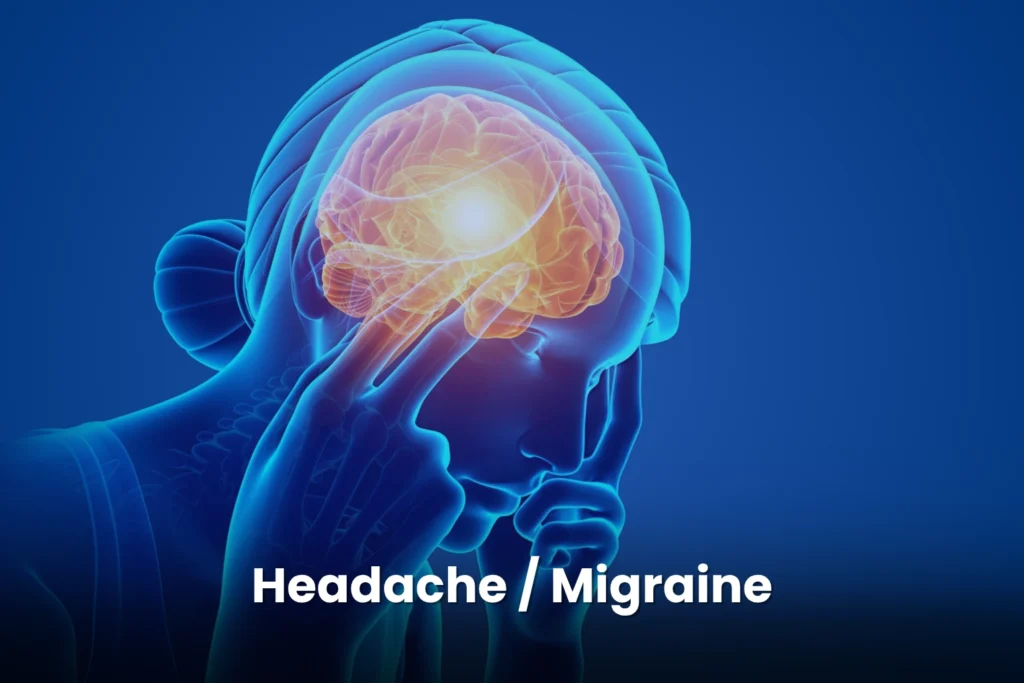
What Is a Migraine? A migraine is a complex neurological disorder marked by repeated episodes of moderate to severe head pain, often throbbing or pulsating. Pain usually affects one side of the head but can occur on both sides and typically lasts from several hours up to three days. Migraines are commonly accompanied by nausea, vomiting, and sensitivity to light, sound, and smell, making daily activities difficult or impossible during an attack. How Do Migraines Affect the Brain? Migraines are more than headaches. They involve changes in nerve signaling, brain chemicals, and blood vessels. Pain is believed to result from inflammation of the dura mater (the membrane between the brain and skull) and activation of pain-sensitive nerves in the brain’s protective layers. These neurological changes explain why migraines cause intense pain along with widespread physical and cognitive symptoms. What Is a Migraine Aura? Some people experience an aura, which consists of temporary neurological symptoms that occur before or during a migraine attack. Aura may include: Visual disturbances (flashing lights, zigzag lines, blind spots) Tingling or numbness in the face or limbs Speech or language difficulties Not everyone with migraines experiences aura, and its presence varies between individuals. What Symptoms Occur Beyond Head Pain? Migraines often cause symptoms beyond head pain, including: Mood changes Extreme fatigue Brain fog Digestive issues Certain rare types, such as hemiplegic migraine, can cause temporary weakness or paralysis on one side of the body, which can mimic a stroke but is usually reversible. What Is the Difference Between Episodic and Chronic Migraine? Migraine is a long-term neurological condition that can be episodic or chronic: Episodic migraine: Fewer headache days per month Chronic migraine: 15 or more headache days per month, with at least 8 having migraine features Chronic migraine significantly affects quality of life and daily functioning. What Are the Most Common Migraine Symptoms? Common migraine symptoms include: Intense, throbbing head pain (usually one-sided) Nausea and vomiting Sensitivity to light (photophobia) Sensitivity to sound (phonophobia) Sensitivity to smells (osmophobia) Pain typically lasts 4–72 hours and can interfere with work, school, and daily life. What Symptoms Can Occur Before, During, or After a Migraine? Additional symptoms may appear at different stages of a migraine attack: Mood changes Difficulty concentrating Fatigue Frequent yawning Food cravings Some individuals experience aura symptoms such as visual changes, tingling, or speech difficulty. What Other Body-Wide Symptoms Can Migraines Cause? Migraines may also cause: Dizziness or vertigo Neck and shoulder pain Sinus pressure or ear pain Increased urination Sensitivity to touch or temperature Gastrointestinal symptoms (stomach pain, diarrhea, constipation) Symptom combinations and severity vary widely between individuals. What Are the Phases of a Migraine Attack? Migraine symptoms often occur in phases: Prodrome: Early warning signs (fatigue, mood changes, food cravings) Aura: Temporary neurological symptoms (when present) Headache phase: Severe pain and associated symptoms Postdrome: Lingering fatigue, brain fog, or mood changes Not everyone experiences every phase. What Types of Migraines Exist? 1. Migraine Without Aura The most common type, featuring moderate to severe one-sided headaches lasting 4–72 hours with nausea and light/sound sensitivity. 2. Migraine With Aura Includes temporary neurological symptoms such as visual, sensory, or speech disturbances before or during the headache. 3. Chronic Migraine Headaches on 15 or more days per month, with at least 8 migraine days. 4. Vestibular Migraine Characterized mainly by dizziness, vertigo, and balance problems, with or without head pain. 5. Hemiplegic Migraine A rare migraine with aura causing temporary weakness or paralysis on one side of the body. 6. Migraine With Brainstem Aura Includes brainstem symptoms such as vertigo, slurred speech, and double vision—without motor weakness. 7. Abdominal Migraine More common in children, causing abdominal pain, nausea, and vomiting. 8. Menstrual Migraine Triggered by estrogen fluctuations during the menstrual cycle. Learn More: What Is Ocular Migraine? Vision Changes & Migraine Types What Causes Migraines? Migraines result from a complex interaction of neurological, vascular, genetic, and environmental factors. A key mechanism is nerve inflammation in the dura mater, leading to altered pain signaling and blood flow. Genetics plays a major role, as migraines often run in families. Learn More: Migraines Explained: Causes, Triggers, and Neurology Care What Are Common Migraine Triggers? Hormonal & Lifestyle Triggers Menstrual cycle, pregnancy, menopause Stress Irregular sleep Skipping meals Physical exertion Sensory & Environmental Triggers Bright or flickering lights Loud noises Strong smells Weather or barometric pressure changes Dietary Triggers Alcohol (especially wine) Caffeine Aged cheeses Processed foods Artificial additives Often, multiple triggers combine to provoke an attack. How Are Neck, Jaw, and Nerves Related to Migraines? Neck-, jaw-, and nerve-related migraines often involve the trigeminal nerve, which supplies sensation to the face, jaw, and head and connects with blood vessels and meninges. Irritation of this nerve can cause pain in the eyes, temples, jaw, or neck. How Are TMJ Disorders Linked to Migraines? TMJ disorders can trigger migraines because the trigeminal nerve supplies the jaw. TMJ inflammation or dysfunction can: Cause jaw or ear pain Trigger migraines Increase migraine frequency through central sensitization How Does Neck Pain Contribute to Migraines? Cervical nerves (C2–C4) connect with the trigeminal nerve at the brainstem, allowing pain signals to overlap. Poor posture, muscle strain, or cervical spine issues can worsen migraines or mimic conditions like cervicogenic headache or occipital neuralgia. What Treatment Options Are Available for Migraines? Migraine treatment focuses on stopping attacks and preventing future episodes. What Medications Treat Acute Migraines? NSAIDs (ibuprofen, aspirin) Acetaminophen Triptans Gepants Anti-nausea medications What Medications Prevent Migraines? Beta-blockers Antidepressants Anticonvulsants CGRP inhibitors How Does Botox Treat Chronic Migraine? Botox is FDA-approved for chronic migraine and involves 31 injections every 12 weeks. It blocks pain signaling and reduces migraine frequency by approximately 50% in responders, with effects typically starting within 2–3 weeks. What Is Infusion Therapy for Migraines? Infusion therapy delivers medication intravenously for severe or refractory migraines. Options include: DHE Ketorolac Antiemetics Preventive infusions like eptinezumab (Vyepti) are given every 12 weeks. When Do Migraines Become a
Head Injury & Concussion Care | Early Neurology Evaluation
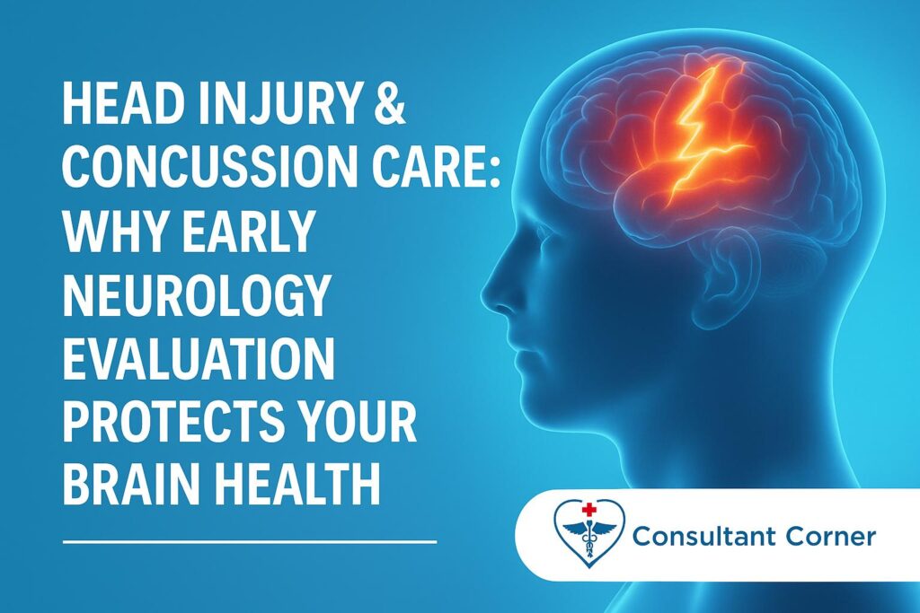
Head Injury & Concussion Care Head injuries are far more common than most people realize—and even what seems like a “mild” injury can disrupt brain function, trigger neurological symptoms, and impact long-term cognitive and emotional health. Whether your head injury came from a car accident, fall, sports impact, workplace incident, or even a minor bump, symptoms may appear immediately or develop over hours or days. Because head injuries are unpredictable, early evaluation by a neurologist is critical. At Consultant Corner, our board-certified neurologists specialize in concussion, traumatic brain injury (TBI), and post-concussion syndrome. We provide comprehensive in-clinic and tele-neurology evaluations, recovery planning, and long-term neurological support to ensure every patient heals safely and fully. ⚠️ WARNING SIGNS AFTER A HEAD INJURY Head injuries affect people differently, and symptoms vary widely. Some patients feel “fine” after the incident, while others experience immediate discomfort. Key symptoms to watch for include: • Persistent or worsening headache• Dizziness, vertigo, or imbalance• Nausea or vomiting• Brain fog, slowed thinking, difficulty concentrating• Short-term memory problems or confusion• Sensitivity to light or noise• Blurred or double vision, trouble focusing• Sleep disturbances or extreme fatigue• Irritability, mood swings, or emotional changes Many of these symptoms indicate a concussion or functional brain disruption. Even if imaging appears “normal,” concussion symptoms can still be present because concussions rarely show up on CT or MRI scans. ❗ WHY EARLY NEUROLOGY CARE MATTERS Early neurology involvement after a head injury is crucial because concussions and mild TBIs are common, frequently under-recognized, and often undertreated. Without timely, targeted care, a significant minority of patients go on to develop prolonged or disabling symptoms. A neurologist can: • Diagnose concussion or mild traumatic brain injury• Identify early signs of post-concussion syndrome• Detect internal bleeding risks such as subdural or epidural hematoma• Identify skull fractures or nerve involvement• Evaluate vision, balance, memory, and cognitive deficits• Assess whiplash-related neurological symptoms• Provide medical clearance for return to work, school, or sports Early intervention improves outcomes, shortens recovery time, and reduces the risk of chronic headaches, vertigo, cognitive decline, and emotional instability. 📊 STATS & FACTS: WHY CONCUSSIONS REQUIRE EXPERT CARE The numbers clearly show why early neurological evaluation is essential after a head injury: • In the U.S., there are approximately 2.8 million traumatic brain injury–related emergency department visits, hospitalizations, and deaths each year, with many involving concussions or mild TBIs. • National data suggest around 3% of Americans report a history of traumatic brain injury, affecting both adults and children. • About 1 in 100 children aged 9–10 sustains a concussion in a single year—representing tens of thousands of pediatric cases annually. • While about 90% of concussion symptoms resolve within 10–14 days, an estimated 10–30% of patients develop persistent symptoms lasting weeks or months (post-concussion or persisting symptoms). • Studies show that 14–40% of patients still report post-concussion symptoms at 3 months, highlighting how easily recovery can be delayed without early identification and management. 🧠 HOW THESE FACTS SUPPORT EARLY NEUROLOGY INVOLVEMENT Because millions of TBIs occur each year—and up to one-third of concussion patients may experience lingering headaches, dizziness, cognitive fog, sleep problems, or mood changes—early specialist evaluation plays a critical role. Neurology care helps to: • Identify patients at higher risk for prolonged recovery• Detect complications such as intracranial bleeding• Diagnose and manage post-concussion syndrome early• Coordinate vestibular and vision rehabilitation• Guide safe, graded return to work, school, or sports• Prevent symptoms from becoming chronic or disabling Early neurological care is not just treatment—it’s protection for long-term brain health. 🏥 WHY PATIENTS TRUST CONSULTANT CORNER Our neurology team is highly experienced in evaluating and treating head injuries of all types. Patients choose Consultant Corner for: ✔ Comprehensive concussion & TBI assessment✔ Thorough review of CT/MRI imaging & ER discharge reports✔ Personalized recovery & graded return-to-activity plans✔ Expert management of headaches, dizziness, cognitive fog, and visual problems✔ Treatment for sleep disturbances, mood changes, and persistent symptoms✔ Tele-neurology options for fast, accessible care from home✔ Same-week appointments—because early care matters We provide clear communication, evidence-based care, and step-by-step recovery guidance designed to rebuild neurological stability and restore quality of life. ⚕️ WHAT RECOVERY LOOKS LIKE Neurological recovery is highly individualized. Your customized treatment plan may include: • Cognitive rest and screen-time modifications• Vestibular therapy for dizziness or balance issues• Vision rehabilitation exercises• Headache and migraine-focused therapies• Sleep hygiene optimization and circadian rhythm support• Stress-reduction and symptom-trigger management• Gradual, monitored return to physical activity Our goal is to help you regain clarity, comfort, and confidence as you return to normal life. 🚨 WHEN TO SEEK EMERGENCY CARE Go to the ER or call 911 immediately if you experience: • Loss of consciousness• Severe or sudden “worst headache” of your life• Repeated vomiting• Weakness, numbness, or tingling on one side• Seizure• Difficulty waking up or severe drowsiness• Clear fluid leaking from the nose or ears• Unequal pupils• Significant confusion or abnormal behavior These symptoms may indicate a medical emergency requiring immediate imaging and intervention. 📞 CONTACT CONSULTANT CORNER If you’ve had a recent head injury and don’t feel like yourself, trust your instincts. Your brain is too important to ignore symptoms. 📱 Phone: +1 888 208 2208📧 Email: info@myconsultantcorner.com🌐 Website: https://myconsultantcorner.com Consultant Corner — Expert Neurology Care When It Matters Most.
What Diabetic Neuropathy Is
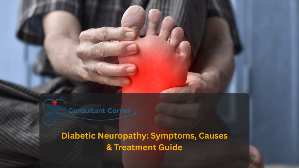
What Diabetic Neuropathy Is Diabetic neuropathy refers to a group of nerve disorders that occur in people with diabetes, affecting sensory, motor, and autonomic nerves. Over time, high glucose levels damage blood vessels and the metabolic pathways that support nerves, leading to progressive dysfunction. The American Diabetes Association describes diabetic neuropathies as the most prevalent chronic complications of diabetes, encompassing diffuse forms like DSPN (symmetrical, length-dependent sensorimotor polyneuropathy due to chronic hyperglycemia and cardiovascular risks) and autonomic neuropathies affecting cardiovascular, gastrointestinal, or urogenital systems. Main Types of Diabetic Neuropathy Peripheral neuropathy:The most common form. It affects the feet and legs first and sometimes the hands, causing pain, tingling, numbness, or loss of sensation in a “stocking-glove” pattern. Autonomic neuropathy:Affects automatic body functions such as heart rate, blood pressure, digestion, bladder control, and sexual function. Proximal (radiculoplexus) neuropathy and focal neuropathies:Causes sudden pain or weakness in specific areas such as the thighs, hips, chest wall, or eye muscles. Common Symptoms Symptoms depend on which nerves are affected, but may include: • Burning, stabbing, or electric-like pain• Numbness or tingling in feet or hands• Reduced ability to feel heat, cold, or injuries• Balance and walking difficulty• Foot ulcers that may not cause pain• Digestive issues such as nausea, bloating, or diarrhea• Dizziness when standing up (orthostatic hypotension)• Bladder control issues• Sexual dysfunction in men and women Loss of protective sensation is especially dangerous because it increases the risk of unnoticed injuries, infections, and amputations. Causes and Risk Factors The primary cause is prolonged high blood sugar, which damages nerves and the small blood vessels that supply them.Additional risk factors include: • Long duration of diabetes• Poor glucose control• High cholesterol• High blood pressure• Kidney disease• Smoking• Obesity• Heavy alcohol use The more risk factors present, the faster nerve damage can progress. Prevention and Self-Care Strategies Preventing or slowing diabetic neuropathy focuses on protecting nerves through comprehensive diabetes management: • Keep blood sugar levels within the target range using medication, nutrition, and physical activity• Maintain healthy cholesterol and blood pressure• Avoid smoking and limit alcohol• Maintain a healthy weight• Perform daily foot checks for cuts, redness, or swelling• Wear comfortable, well-fitting shoes• Schedule regular podiatry visits to prevent ulcers and infections Good foot care is critical to reducing the risk of complications. Treatment Basics Good blood glucose management is the most important treatment to slow nerve damage and may improve symptoms, especially in the early stages.Painful diabetic neuropathy can be managed with: • Certain antidepressants or anticonvulsants• Topical treatments such as capsaicin or lidocaine• Physical therapy• Exercise programs• Non-drug therapies such as TENS (nerve stimulation) Treatment plans are personalized based on symptom severity, medical history, and patient goals. Statistics and Key Facts • Over 50% of people with diabetes develop some form of neuropathy• About 25% experience painful diabetic peripheral neuropathy• Neuropathy increases the risk of foot ulcers and amputations• Early management dramatically reduces complications Quick Reference Guide Aspect Key Points What it is Diabetes-related nerve damage affects peripheral, autonomic, and other nerves Main symptoms Burning or stabbing pain, numbness, tingling, balance issues, and autonomic dysfunction Main causes Long-term high blood sugar plus factors like cholesterol issues, hypertension, obesity, smoking, and kidney disease Prevention focus Safe glucose control, blood pressure, and lipid management, foot care, smoking/alcohol reduction How common it is Over 50% of people with diabetes develop neuropathy; about 25% experience painful neuropathy Telehealth Evaluation for Diabetic Neuropathy in Bloomington, IL. At Consultant Corner, your neurologist provides a complete virtual assessment, including: • Review of symptoms and medical history• Neurological screening by video• Differentiating neuropathy from other nerve disorders• Foot risk assessment and preventive guidance• Medication recommendations for nerve pain• Lifestyle and nutrition strategies that support nerve health• Referrals for additional testing if needed• Ongoing follow-up and treatment adjustments Telehealth makes expert neurological care accessible, convenient, and personalized. When to Seek Urgent Evaluation Seek immediate medical help if you experience: • Worsening weakness or inability to walk• Foot wounds that are not healing• Severe or sudden pain in thighs, hips, or chest• New bladder control problems• Signs of infection in the feet• Rapid progression of numbness These may indicate severe nerve damage or complications that require prompt care. Get Expert Neuropathy Care From Consultant Corner If you are experiencing nerve pain, numbness, tingling, or foot problems related to diabetes, do not wait. Early treatment can prevent serious complications and improve your long-term quality of life. 👉 BOOK TELEHEALTH APPOINTMENT👉 CONTACT CONSULTANT CORNER TODAY
What Is Cervical Radiculopathy?
What Is Cervical Radiculopathy? Cervical radiculopathy happens when one of the cervical spinal nerve roots is irritated or compressed. This often results from cervical disc herniation, degenerative disc disease, cervical spondylosis (arthritis), or foraminal stenosis (narrowing of the nerve root canal). When a nerve root is compressed, inflammation develops, leading to neurological symptoms that follow the exact path of the affected nerve (C5, C6, C7, etc.). This condition is a common cause of neck pain with arm symptoms, especially in adults aged 30–60. According to the National Center for Biotechnology Information, “Neck pain is a common condition that can cause significant discomfort and disability in patients of different ages. Workers who have experienced neck pain account for up to 40% of work absenteeism. Cervical radiculopathy, on the other hand, is a condition in which the nerve root of a spinal nerve is compressed or impaired, causing pain and symptoms to spread beyond the neck and radiate to other areas of the body, such as the arms, neck, chest, upper back, and shoulders. Due to the nerve impingement, muscle weakness and impaired deep tendon reflexes are often observed. Cervical radiculopathy is a common neck disorder that reduces function, productivity, and quality of life. The natural course is generally favorable. However, significant pain, progressive neurologic loss, or myelopathic signs warrant escalation of care.” Common Symptoms of Cervical Radiculopathy Symptoms typically include both neck discomfort and arm-related neurological signs, such as: • Sharp, burning, throbbing, or electric-like pain• Radiating pain into the shoulder, upper arm, forearm, or hand• Numbness or “pins and needles” sensations• Muscle weakness, such as difficulty lifting or gripping objects• Reduced reflexes• Pain that worsens with neck rotation or extension• Relief when resting or supporting the arm These symptoms distinguish radiculopathy from simple mechanical neck pain. What Causes Cervical Radiculopathy? The two leading causes include: Cervical Disc Herniation: More common in younger adults. A bulging or ruptured disc presses on the nerve root. Cervical Spondylosis (Arthritis): Age-related degeneration leads to bone spurs or narrowing of the foramina, compressing the nerve root. Less common causes include trauma, tumors or cysts, congenital narrowing, postural strain, and vascular abnormalities. Risk Factors That Increase Neck Nerve Compression Certain lifestyle and workplace factors significantly increase risk: • Long hours of computer use with poor posture• Repetitive neck flexion or extension• Heavy lifting or jobs that strain the neck• Operating vibrating equipment• Smoking• Obesity• Weakened neck and upper-back muscles These factors accelerate wear and tear on the cervical spine over time. Cervical Radiculopathy vs Simple Neck Pain Aspect Cervical Radiculopathy Simple Mechanical Neck Pain Main issue Pinched/irritated nerve root Muscle or ligament strain Pain pattern Radiates down the arm, often below the elbow Stays in the neck/shoulder Neurological signs Tingling, numbness, weakness, reflex loss Rare Typical causes Disc herniation, arthritis, and foraminal stenosis Poor posture, overuse Severity Can impair daily function Usually mild to moderate How to Help Prevent Cervical Radiculopathy Prevention focuses on spinal health, posture support, and reducing repetitive strain. Improve posture and ergonomics: Keep screens at eye level, avoid hunching, and take short movement breaks every 20–30 minutes. Strengthen neck and upper-back muscles: Stretching, light resistance exercises, and aerobic conditioning help stabilize the spine. Use safe lifting techniques: Bend at the hips and knees, keep loads close to your body, and avoid overhead lifting when possible. Optimize sleep position: Use a supportive pillow, maintain a neutral neck posture, and avoid extreme rotation during sleep. Key Facts & Statistics About Cervical Radiculopathy Population studies show: • Affects 80–85 people per 100,000 each year• Incidence in men is approximately 107 per 100,000• Incidence in women is around 63 per 100,000• Most commonly occurs in people aged 40–55• The C7 nerve root is affected in over 50% of cases• The C6 nerve root is involved in about 25% of cases These numbers demonstrate how common cervical nerve compression is among adults with spinal degeneration or neck strain. When Cervical Radiculopathy Requires Urgent Evaluation Seek immediate medical attention if you experience: • New or worsening arm weakness• Difficulty walking or balancing• Loss of coordination• Sudden, severe neck pain• Bladder or bowel changes• Pain that does not improve with rest• Symptoms following trauma or injury These can indicate severe nerve or spinal cord compression. Telehealth Evaluation for Cervical Radiculopathy Through telehealth, your neurologist can perform a thorough virtual assessment, including: • Detailed symptom evaluation• Neurological screening• Differentiating radiculopathy from other causes• Medication recommendations• Physical therapy and posture guidance• Imaging referrals (MRI, CT, X-ray)• Follow-up monitoring and adjustments Early treatment leads to faster recovery and prevents long-term complications.
Neurological Disorders Affect Half of Americans
Neurological Disorders Now Affect Over Half of Americans, New Research Shows Abstract Neurological and nervous-system disorders are a major source of disability in the United States. GBD 2021 data show 180.3 million Americans (54.2%) had at least one nervous system disorder in 2021. The analysis covered 36 conditions, including neurological, congenital, systemic, and infectious disorders. These conditions caused 16.6 million DALYs nationwide. The greatest disability burden came from stroke, Alzheimer’s and dementias, diabetic neuropathy, and migraine. Prevalence was highest among adults 80 years and older. State-level DALY rates varied widely. Since 1990, prevalence has stayed stable, mortality has declined, and YLDs have increased, indicating more chronic disability. Findings emphasize the need for better research, stronger public health planning, and improved resources to address the rising neurological burden in the U.S. Neurological and nervous-system–related disorders have become one of the most significant contributors to disability in the United States, according to new findings published in JAMA Neurology. Researchers report that more than 54% of the U.S. population — approximately 180 million people — live with at least one condition affecting the brain, spinal cord, or peripheral nerves. Lead investigator Dr. John P. Ney of Yale University explained that the burden extends well beyond classic neurological illnesses. Many systemic and developmental conditions, including diabetic neuropathy, autism spectrum disorders, and metabolic diseases, also play a major role in shaping nervous system health. Key Data From the Global Burden of Disease (GBD) 2021 Analysis Using cross-sectional U.S. data from 1990–2021, researchers evaluated 36 nervous system–related disorders, including neurological, congenital, neonatal, infectious, and systemic conditions. The study assessed disability-adjusted life-years (DALYs), years lived with disability (YLDs), years of life lost (YLLs), prevalence, and state-level variations. Most Common Nervous-System Conditions in 2021 Tension-type headache: 121.9 million people Migraine: 57.7 million people Diabetic neuropathy: 17.1 million people These conditions are major drivers of chronic disability and reduced productivity. Leading Causes of Neurological Disability Stroke: 3.9 million DALYs Alzheimer’s disease & dementias: 3.3 million DALYs Diabetic neuropathy: 2.2 million DALYs Migraine disorders: 2.1 million DALYs Stroke and dementia remain dominant, but neuropathy and chronic headache disorders contribute heavily to daily functional impairment. Geographic Differences in Neurological Health Burden Highest DALY rates were recorded in: Mississippi Missouri Alabama Indiana Iowa Louisiana South Carolina Lowest rates were found in: California New York Washington New Jersey Age and Sex Trends Adults 80+ years had the highest DALY rates (32,928). Children under 5 years experienced a higher burden than those aged 5–19. Burden increased 2x–4x across adult age categories. Women showed slightly higher prevalence, but men had more deaths and YLLs. Long-Term Trend (1990–2021) Overall prevalence remained stable Mortality declined 14.6% YLDs increased 9.8%, indicating more people survive but live with chronic impairment Dr. Ney emphasized that understanding this expanding burden is essential for shaping U.S. research funding, neurotherapeutic development, and clinical resource allocation. Expert Neurological Care at My Consultant Corner My Consultant Corner is a trusted neurology clinic serving patients in Bloomington, IL. The practice is dedicated to providing compassionate, evidence-based neurological care through both in-person visits and convenient telehealth neurology services. Whether you’re dealing with migraines, neuropathy, cognitive concerns, or long-term neurological conditions, their specialist team ensures timely assessment, personalized treatment plans, and ongoing support to improve your quality of life. Take the Next Step Toward Better Neurological Health If you need reliable neurological services in Bloomington—or prefer flexible care from home—My Consultant Corner is here to help.👉 Schedule your neurology or telehealth appointment today and get the expert care you deserve.
2025’s Final Supermoon Arrives Tonight — What Makes the Cold Moon Special

December’s Cold Moon: How to Watch the Last Supermoon of 2025 Key Points December’s Cold Moon is the last supermoon of 2025. It will rise on Thursday, December 4, appearing larger and brighter. A supermoon occurs when the full Moon aligns with perigee, its closest point to Earth. UK moonrise times range from 14:29 to 15:05 GMT, depending on the city. The Cold Moon will appear near the Pleiades, Aldebaran, Jupiter, and Orion. Early clouds may affect parts of the UK, but clear skies are expected overnight. December’s full Moon is traditionally called the Cold Moon, Long Night Moon, and Moon Before Yule. The event aligns with anniversaries of Apollo 8 and Apollo 17. The Moon rises during a Major Lunar Standstill, giving it a higher and more northern path. Viewers can watch live online via the Virtual Telescope Project and Griffith Observatory. Cold Moon 2025: Exact Time, Weather Forecast, and How to Watch Live Stargazers have one final lunar spectacle to look forward to in 2025. December’s Cold Moon, which also happens to be the year’s final supermoon, will rise on Thursday, 4 December, lighting up the sky with an unusually bright and enlarged glow. This event marks the third consecutive supermoon of the year — a sequence that won’t occur again until early 2026. What Makes the Cold Moon a Supermoon? The Moon becomes a supermoon when it reaches perigee, the closest point in its elliptical orbit around Earth. If that moment coincides with the full Moon phase — an alignment astronomers call syzygy — the Moon appears noticeably bigger and shines more intensely. While supermoons are relatively common, appearing several times a year, seeing three back-to-back adds an extra layer of rarity to this December event. Exact Time to Spot the Cold Moon On 4 December, the Cold Moon will begin its ascent in the northeastern sky, offering skywatchers across the UK a long viewing window. Moonrise Schedule London: 14:52 GMT Edinburgh: 14:29 GMT Belfast: 14:52 GMT Cardiff: 15:05 GMT The Moon will remain visible until around 8–9 a.m. the next morning, giving plenty of time for night photography or casual viewing. Night-Sky Companions As it climbs higher, the Cold Moon will appear near: The shimmering Pleiades The red star Aldebaran Brilliant Jupiter The winter constellation Orion This natural lineup creates ideal conditions for both stargazers and photographers hoping to capture a dramatic scene. Weather Outlook for Viewing A band of cloud and rain is forecast to push eastward across parts of the UK on Thursday. Northern and eastern regions may experience reduced visibility early in the evening. However, skies are expected to clear overnight, with misty patches and frosty temperatures developing in some locations. Checking your local weather report before heading outside is recommended. Why December’s Full Moon Is Called the “Cold Moon” The name “Cold Moon” has deep roots in traditional seasonal calendars. Long before modern timekeeping, Indigenous cultures and early societies named each Moon based on seasonal cues. December’s Moon is associated with: Cold Moon — marking winter’s arrival Long Night Moon — reflecting the lengthy winter nights Moon Before Yule — tied to pre-solstice traditions Snow Moon (Cherokee) Winter Maker Moon (Abenaki) This full Moon appears just weeks before the winter solstice, the darkest and shortest day of the year. A Celestial Event with NASA History December’s full Moon also aligns with significant anniversaries from NASA’s lunar program. Apollo 8 Launched on 21 December 1968, it was the first crewed mission to orbit the Moon. Apollo 17 Lifted off on 7 December 1972, marking humanity’s last landing on the lunar surface. NASA’s Noah Petro, project scientist for Artemis III, notes that the Cold Moon offers a meaningful opportunity to reflect on past exploration and the missions ahead. The Cold Moon and the Future of Artemis This final supermoon appears only months before the scheduled launch window of Artemis II, expected between February and April 2026. The mission will send astronauts on a journey around the Moon — the first human trip beyond low Earth orbit in more than five decades. Petro says the Cold Moon is a fitting reminder of how close humanity is to returning astronauts to deep space. How to Watch the Supermoon Online If clouds or city lights limit your view, several livestreams will broadcast the Cold Moon in real time. 🌕 Virtual Telescope Project (Italy) Dec 4 at 3 p.m. EST / 20:00 GMTAstronomer Gianluca Masi will share live telescopic visuals from Italy, along with some of his best lunar photos. 🌕 Griffith Observatory (Los Angeles) Dec 5 at 8:27 p.m. EST / 01:27 GMT (Dec 6)Watch the Moon rise over Los Angeles from the iconic observatory on Mount Hollywood. A Rare Alignment: The Major Lunar Standstill December’s Cold Moon rises during the final stage of a Major Lunar Standstill, a rare 18.6-year cycle when the Moon’s orbit tilts at its maximum angle. This makes the Moon: Rise farther north Reach a higher altitude Follow a more dramatic path across the sky It’s one of the most photogenic lunar moments of the decade. Tips for the Best Viewing Experience To make the most of the Cold Moon: Choose a spot with an open view of the eastern horizon Bring binoculars or a telescope for enhanced detail Photograph the Moon during moonrise for the largest apparent size Compare live views with livestreams for a richer experience Bottom Line December’s Cold Moon isn’t just the last full Moon of the year — it’s the final supermoon of 2025, arriving with astronomical significance, cultural meaning, and historical ties to NASA’s lunar missions. Whether you catch it in person or through online broadcasts, this supermoon is one of the year’s most captivating astronomical events.
Daily Orange Juice May Reduce Inflammation, Study Finds

A Daily Glass of Orange Juice May Reduce Inflammation and Support Heart Health, New 2025 Study Finds A new 2025 study is reshaping what we know about orange juice. Scientists say a daily glass of pure orange juice may do far more than boost energy — it may help lower inflammation, improve blood pressure regulation, and support long-term heart health. A Simple Drink With a Big Impact Researchers asked adults to drink 500 ml of pasteurised orange juice every day for 60 days. The results surprised them: thousands of genes inside immune cells changed their activity. Several key inflammation-related genes — NAMPT, IL6, IL1B, NLRP3 — became less active. Another gene, SGK1, which influences sodium retention and blood pressure, also quieted down. These changes align with earlier studies showing orange juice can gently reduce blood pressure in young adults. Why Orange Juice Works Experts point to hesperidin, a natural citrus flavonoid with antioxidant and anti-inflammatory properties. It supports: Blood vessel relaxation Cholesterol balance Healthier sugar metabolism This may explain why orange juice has shown consistent heart-health benefits across studies. Metabolism, Weight, and Gut Health The response isn’t the same for everyone. Overweight participants showed larger changes in fat-metabolism genes. Lean participants experienced stronger reductions in inflammation genes. Other research adds to the picture. In 639 people across 15 trials, orange juice lowered insulin resistance and LDL cholesterol. Studies also show it may: Improve endothelial function Support healthy gut bacteria Reduce inflammatory markers Improve blood flow Even small improvements in these areas can add up over years. More Than a Breakfast Drink Orange juice is part of daily routines and cultural traditions around the world — and now science suggests it may also serve as a functional food offering measurable cardiovascular benefits. Researchers emphasize that whole fruit is still the best option due to fibre, but a modest daily serving of pure orange juice appears to support: Lower inflammation Better blood pressure control Improved metabolic health A simple, familiar drink may be more powerful than we ever imagined. references: https://theconversation.com/your-daily-orange-juice-could-be-helping-your-heart-270492 https://www.euronews.com/health/2025/12/02/orange-juice-found-to-affect-thousands-of-genes-in-immune-cells-study-says
can cervical radiculopathy cause headaches

Can cervical radiculopathy cause headaches? Yes. Cervical radiculopathy can be associated with headaches, often called cervicogenic headaches when the pain is referred from the neck into the head. How it causes headaches? When a nerve root in the cervical spine is compressed or irritated (from a disc herniation, arthritis, or foraminal narrowing), pain signals can travel along the nerve’s pathway and be perceived in the head as a headache. This is especially recognized with involvement of the upper cervical levels (roughly C1–C4), which share connections with nerves that supply sensation to parts of the head and face. Typical headache features Headaches linked to cervical radiculopathy or other neck pathology often: Start in the back of the neck or base of the skull and radiate upward into the head. Are more on one side, usually the same side as the neck and arm symptoms. May be accompanied by neck pain, reduced neck motion, and arm pain, tingling, or weakness When to seek evaluation? Red flags that should prompt prompt medical evaluation include: New or worsening severe headache, especially with neurological symptoms (weakness, trouble speaking, vision changes, balance problems, confusion). Headache after trauma, fever or stiff neck, or a sudden “worst headache of life.” Common Symptoms of Cervicogenic Headaches in Cervical Radiculopathy If you’re experiencing any of the following symptoms, your headaches may be originating from your cervical spine rather than the head itself: Pain that begins at the base of the skull or back of the neck and then radiates upward Head pain on one side, often matching the side of neck or arm discomfort Neck stiffness or reduced ability to turn or move the neck Arm pain, tingling, numbness, or weakness, especially when a cervical nerve root is irritated Pain that worsens with neck movement, posture, or prolonged positioning These features help distinguish cervicogenic headaches from migraines, tension headaches, or sinus-related headaches, which typically have different patterns. When to Seek Medical Evaluation Headaches linked to cervical issues are often treatable, but certain warning signs mean you should seek medical evaluation promptly. Get checked if you experience: A sudden, severe headache unlike any you’ve had before Headache accompanied by weakness, vision changes, speech difficulty, or balance problems Headache with fever, neck stiffness, or occurring after an injury or accident Persistent headaches that do not improve with rest or common medications Neck pain that radiates into the shoulder, arm, or hand Many people delay seeking care until symptoms worsen, but early evaluation helps pinpoint the cause and leads to more effective treatment. Treatment Options for Cervical Radiculopathy–Related Headaches Individuals experiencing headaches linked to cervical radiculopathy may benefit from several treatment approaches designed to reduce nerve irritation, improve mobility, and relieve pain. 1. Physical Therapy Targeted exercises can help improve posture, strengthen cervical muscles, and reduce pressure on irritated nerves. 2. Posture Correction & Ergonomics Adjusting daily posture and optimizing work or home ergonomics are essential, especially for people who sit for long periods or engage in repetitive neck movements. 3. Medications Anti-inflammatory drugs, muscle relaxants, or neuropathic pain medications may be used to manage pain and decrease nerve irritation. 4. Interventional Procedures For more persistent or severe symptoms, options such as nerve blocks or epidural steroid injections may provide relief and reduce inflammation around the affected cervical nerve root. 5. Telehealth Support Many individuals now choose virtual consultations for convenience, follow-up care, and faster access to healthcare guidance, especially when managing chronic neck or nerve-related symptoms. How to differentiate cervicogenic headache from migraine? Cervicogenic headache and migraine can look similar, but they differ in what triggers them, associated neck findings, and classic migraine features. Key clinical differences Cervicogenic headache usually starts in the neck or back of the head and is clearly linked to neck movement, sustained posture, or pressure over specific cervical/occipital areas; the neck is often stiff and tender, with reduced range of motion. Migraine usually starts in the head (often in the frontotemporal region or around one eye), are not consistently triggered by neck movement, and often have a pulsating or throbbing quality. Laterality and pain quality Cervicogenic headache is typically one‑sided, on the same side as neck or shoulder/arm pain, and the pain is often steady, non‑throbbing, and can radiate from the neck upward in a “ram’s horn” pattern. Migraine is also commonly one‑sided but may switch sides between attacks and is classically throbbing, moderate to severe, and worsened by routine physical activity. Neurologic and sensory symptoms Cervicogenic headache usually lacks migraine aura, prominent nausea, or strong light and sound sensitivity; when these occur, they are typically milder and secondary to pain rather than defining features. Migraine often includes nausea, vomiting, photophobia, and phonophobia, and some patients have aura (visual or sensory disturbances such as flashing lights, zigzags, or numbness) developing before or with the headache. Physical exam and diagnostic clues In cervicogenic headache, examination often shows a reproducible pattern of cervical musculoskeletal impairment: reduced neck movement, painful or stiff upper cervical joints, and muscle dysfunction; symptoms may be provoked by neck tests or pressure on facet joint/occipital regions, and may improve with diagnostic cervical nerve or joint blocks. In migraine, the neck exam may be normal or show only nonspecific tenderness; diagnosis is clinical, using headache history that meets International Classification of Headache Disorders criteria (recurrent attacks, duration 4–72 hours, pulsating quality, associated nausea or photophobia/phonophobia, etc.). Practical table: cervicogenic vs migraine Feature Cervicogenic headache Migraine headache Primary source Neck (joints, discs, soft tissues) Primary brain-based disorder Onset/location Begins in neck/occiput, spreads to head Begins in head (often frontotemporal/periorbital) Triggered by Neck movement, posture, or palpation Stress, hormones, certain foods, sleep, stimuli Pain quality Steady, dull, non‑throbbing usually Pulsating/throbbing common Associated neck signs Marked stiffness, reduced ROM, focal tenderness Neck may feel tight but exam often nonspecific Aura Absent Often present in migraine with aura Nausea/photophobia Uncommon or mild Common and often prominent Diagnostic confirmation Response to cervical blocks/targeted PT Clinical criteria; no specific physical trigger If your headaches change with neck movement, posture, or pressure
What Causes Alzheimer’s Disease?

What Causes Alzheimer’s Disease? Alzheimer’s disease is a complex condition with multiple contributing factors. It doesn’t arise from a single cause, but rather from a combination of abnormal brain changes and influences from genetics, health, and lifestyle factors over an extended period. Brain Changes in Alzheimer’s In Alzheimer’s disease, two key proteins, beta-amyloid and tau, undergo abnormal changes in the brain. Beta-amyloid forms sticky clumps, known as plaques, which accumulate between neurons (nerve cells). Tau, on the other hand, creates tangles inside neurons. These plaques and tangles disrupt normal communication between brain cells, depriving them of essential nutrients, ultimately leading to cell death and the shrinkage of the brain over time. 7 Common Signs of Alzheimer’s Disease The early signs of Alzheimer’s can be subtle and might initially be mistaken for typical aging. However, as the disease progresses, these symptoms become more noticeable and affect daily life: Memory loss that disrupts daily life: For example, forgetting important dates or repeatedly asking for the same information. Challenges in planning or problem-solving: This may include difficulty concentrating or managing numbers (e.g., balancing a checkbook). Difficulty completing familiar tasks: Routine tasks at home, work, or leisure (such as driving to a familiar location) can become hard to manage. Confusion with time or place: A person might lose track of dates, seasons, or even forget where they are. Trouble understanding visual images and spatial relationships: This can impact the ability to read, judge distance, or recognize faces. Problems with language: Difficulty finding the right words or following and participating in conversations. Changes in mood and personality: Individuals may become more depressed, withdrawn, or irritable. There may also be mood swings, aggression, or loss of inhibitions. These signs typically worsen over time and can severely impact the person’s daily functioning and independence. When to Seek a Doctor It’s important to seek a doctor if someone shows multiple signs of Alzheimer’s, especially if the symptoms: Are noticeable and worsening over time Interfere with daily life and routine activities Include memory loss, confusion, trouble completing familiar tasks, or changes in personality or behavior Cause concern for the individual or their family members Early evaluation by a healthcare professional is recommended because prompt diagnosis can help manage symptoms, rule out other causes, and provide support and planning options. Underlying Causes and Risk Factors Alzheimer’s is thought to develop due to a mix of factors, including age-related brain changes, genetic predispositions, cardiovascular health, and lifestyle choices. While no single factor causes Alzheimer’s, certain risk factors significantly increase the likelihood of developing the disease: Age: The greatest known risk factor. Alzheimer’s typically affects those over 65, with risk increasing significantly as individuals age. Genetics: Variants of the APOE ε4 gene are strongly linked to a higher risk of Alzheimer’s. Family History: Having close relatives with Alzheimer’s increases the risk. Down Syndrome: Individuals with Down syndrome are at higher risk due to an extra copy of chromosome 21. Traumatic Brain Injury: Past head injuries are associated with a higher risk of Alzheimer’s. Lifestyle Factors: Factors such as heavy alcohol use, poor sleep, smoking, and obesity are linked to increased risk. Chronic Health Conditions: Conditions like high blood pressure, high cholesterol, and type 2 diabetes (especially if poorly controlled) increase vulnerability. Environmental Factors: Exposure to air pollution is also considered a potential risk factor. Hereditary vs. Sporadic Alzheimer’s The majority of Alzheimer’s cases are sporadic, meaning the disease arises due to a combination of genetic, environmental, and lifestyle factors. These cases don’t have a guaranteed genetic link and typically develop later in life. However, around 1–2% of Alzheimer’s cases are genetic and due to rare inherited mutations in specific genes such as APP, PSEN1, and PSEN2. These cases are often linked to early-onset Alzheimer’s, typically occurring before the age of 65. Known and Unknown Aspects of Alzheimer’s Research indicates that protein buildup, inflammation, and vascular problems (related to blood vessels) begin many years—sometimes decades—before symptoms like memory loss or cognitive decline emerge. However, the exact reasons why these abnormal protein changes occur in one person and not another are not fully understood. Additionally, how various risk factors interact in each individual is still an area of ongoing research. Can Alzheimer’s Risk Be Reduced? While no method is proven to completely prevent Alzheimer’s, certain lifestyle changes can lower the risk of developing the disease: Heart and vascular health: Maintaining a healthy heart and blood vessels is crucial for brain health. Physical activity: Regular exercise has been shown to have protective effects on the brain. No smoking: Smoking accelerates brain aging and contributes to Alzheimer’s risk. Moderate alcohol consumption: Limiting alcohol intake is linked to a lower risk of cognitive decline. Healthy diet: Following a Mediterranean-style diet, rich in fruits, vegetables, and healthy fats, may help reduce Alzheimer’s risk. Good sleep: Adequate and restful sleep is essential for maintaining cognitive function. Mental and social engagement: Staying intellectually and socially active is linked to a reduced risk of dementia. Although these measures are associated with a lower risk of Alzheimer’s, it’s important to note that they cannot guarantee prevention. Conclusion Alzheimer’s disease is a multifactorial condition, influenced by both genetic and environmental factors. Although much is still unknown about why some people develop Alzheimer’s and others do not, lifestyle modifications aimed at improving heart and brain health may significantly reduce the risk. Regular medical checkups and early intervention remain vital for managing the disease and supporting brain health throughout life. Seek Expert Alzheimer’s Care in Bloomington, IL If you or a loved one is experiencing symptoms of Alzheimer’s disease, early intervention is key. At My Consultant Corner, we specialize in Alzheimer’s treatment and management in Bloomington, IL, providing comprehensive care to help manage symptoms and improve quality of life. Our team of neurological experts is dedicated to offering personalized care plans, ensuring that patients receive the support they need. Don’t wait — schedule an appointment with our specialists today and take the first step toward
5 Ways to Improve Mental Health Naturally & Effectively

5 Proven Ways to Improve Mental Health Naturally Mental health is a core part of overall well-being, yet it’s often overlooked in the busy routines of daily life. The good news is this: you don’t need drastic changes to support your mind. Small, evidence-backed daily habits can dramatically improve emotional balance, reduce stress, and build long-term resilience. In this guide, we explore five science-supported ways to improve mental health—including physical activity, mindfulness, sleep habits, nutrition, social connections, and daily practices that nurture emotional well-being. Why Daily Habits Matter for Mental Health Mental health is shaped by lifestyle, environment, relationships, biology, and daily routines. Research consistently shows that building healthy habits can: Reduce anxiety and stress Boost mood and happiness Strengthen emotional regulation Support cognitive function Improve overall life satisfaction These small habits don’t replace therapy or medical care, but they make excellent foundational practices that support better mental well-being. 1. Build and Strengthen Social Connections Human connection is one of the strongest protective factors for mental health. People with meaningful social bonds experience less stress, better emotional stability, and stronger feelings of belonging. Why Social Connection Supports Mental Health Reduces loneliness and isolation Boosts emotional support Strengthens self-esteem Improves resilience during tough times Ways to Strengthen Social Relationships Talk to family or friends regularly Join a club, community group, or interest class Stay connected through shared activities Reach out when you’re feeling overwhelmed Even one supportive relationship can make a significant difference. 2. Stay Physically Active for a Healthier Mind Exercise is one of the most reliable natural antidepressants. Physical activity releases endorphins—your brain’s “feel-good chemicals”—while reducing cortisol, the stress hormone. Mental Health Benefits of Exercise Improves mood and emotional balance Reduces anxiety and stress Enhances sleep quality Increases confidence and energy levels Simple Activities to Start With 20–30 minutes of brisk walking Yoga or stretching Light home workouts Dancing, cycling, and swimming Consistency matters more than intensity. 3. Practice Mindfulness and Present-Moment Awareness Mindfulness is the practice of focusing on the present moment without judgment. This simple habit helps reduce overthinking, improve emotional regulation, and create a sense of calm. Why Mindfulness Helps Reduces stress and tension Improves focus and clarity Lowers anxiety and overwhelm Helps manage negative thoughts Easy Mindfulness Practices Guided meditation (5 minutes a day) Deep breathing exercises Body-scan relaxation Journaling your thoughts Mindful walking Mindfulness trains the brain to respond calmly rather than react automatically. 4. Engage in Acts of Kindness and Gratitude Giving to others is linked to greater happiness, emotional well-being, and improved mental health. Acts of kindness activate areas of the brain associated with pleasure and reward. Benefits of Kindness Increases positive emotions Strengthens social bonds Improves self-worth Reduces stress Simple Ways to Practice Kindness Help a friend or neighbor Donate or volunteer Write a message of appreciation Perform a small anonymous act of kindness Gratitude also shifts focus from problems to the positives in life. 5. Prioritize Quality Sleep and Healthy Nutrition Sleep and nutrition directly influence the brain, mood, and emotional stability. Poor sleep or nutrient deficiencies often worsen symptoms of anxiety or depression. Why Sleep Matters Supports mood regulation Improves memory and focus Reduces irritability and low motivation Sleep Hygiene Tips Maintain a consistent sleep schedule Limit screens before bed Create a relaxing bedtime routine Nutrition for Mental Health Include omega-3 fatty acids (salmon, walnuts) Eat fresh fruits and vegetables Choose whole grains Limit sugar and processed foods Nourishing your body also nourishes your mind. Long-Term Benefits of Healthy Mental Health Habits By incorporating these simple actions daily, you can experience: Greater emotional stability Better stress tolerance Higher energy and motivation Improved relationships Stronger overall well-being Mental wellness is built through steady, consistent habits—not perfection. Reference: https://www.nhs.uk/mental-health/self-help/guides-tools-and-activities/five-steps-to-mental-wellbeing/ FAQ What are the best daily habits for improving mental health? Regular exercise, mindfulness practice, quality sleep, healthy nutrition, social connection, and acts of kindness are among the most effective evidence-based habits for improving mental health. How does exercise help mental health? Physical activity releases endorphins, reduces stress hormones, improves mood, and supports better sleep, making it one of the most effective natural ways to improve mental well-being. Why is sleep important for mental health? Good sleep helps regulate mood, reduce anxiety, improve focus, and support emotional resilience. Poor sleep is strongly associated with higher stress and depressive symptoms. Can mindfulness reduce anxiety? Yes. Mindfulness and deep-breathing practices reduce stress, calm racing thoughts, and improve the brain’s ability to handle anxiety. Does helping others improve mental health? Acts of kindness and gratitude release positive brain chemicals, improve mood, and create a sense of purpose—boosting emotional well-being.
Cervical Radiculopathy & Neck Pain
⚡ Cervical Radiculopathy & Neck Pain Neck Pain With Arm Numbness or Tingling? You May Have a Pinched Nerve — We Can Help. A pinched nerve in the neck (cervical radiculopathy) can cause neck pain that shoots into the shoulder, arm, or hand. Patients often describe burning, numbness, tingling, electric pain, or weakness — symptoms that rarely improve without proper neurological care. At Consultant Corner, we specialize in diagnosing and treating nerve-related neck pain so you can get relief fast. 👉 If your neck pain travels down your arm, it’s time for a specialist. ⚠️ Key Symptoms of Cervical Radiculopathy 🟣 Neck pain radiating to shoulder or arm 🔵 Numbness, tingling, or “pins and needles” 🟢 Burning or electric shocks down the arm 🟠 Weakness in the arm or hand 🟡 Pain worse with head turning or looking down 🔺 Difficulty gripping or dropping objects These symptoms point to compression of the C5–C8 nerves — and often require targeted treatment. 🔍 Common Causes (SEO-Friendly Terms) Herniated cervical disc Bulging disc in neck Cervical spinal stenosis Degenerative disc disease Arthritis of the neck Tech neck / posture strain Nerve compression from muscle spasm Search engines favor these exact terms — and so do your patients. 🏥 Why Patients Choose Consultant Corner This is where your differentiators matter. Patients want fast answers and real relief. 🧠 ✓ Neurology Expertise in Pinched Nerves We identify which nerve is affected — and why. 💥 ✓ Targeted, Non-Surgical Treatment Options Nerve pain medications, muscle relaxation strategies, posture correction, ergonomic planning. 📉 ✓ Imaging Review by Neurology We interpret MRIs, CTs, and X-rays to create a clear, actionable plan. 🎯 ✓ Personalized Recovery Strategy Reduce pain, restore movement, prevent long-term nerve damage. ⏱️ ✓ Same-Week Appointments Because nerve pain should never wait. 👉 Our goal: fast relief, long-term stability, and preventing permanent nerve injury. 🚨 When Neck Pain Requires Urgent Care Seek immediate evaluation if you experience: Sudden weakness New numbness spreading down the arm Loss of balance or coordination Loss of bladder/bowel control Severe neck pain after trauma These may indicate a serious spinal issue. 📞 Get Relief From Neck & Arm Pain Today Cervical radiculopathy is highly treatable, especially when caught early.Don’t wait for numbness or weakness to become permanent. 👉 Book Your Neck Pain / Pinched Nerve Evaluation 📞 Call or Request Online
Diabetic Neuropathy (Nerve Damage)
🦶🔥 Diabetic Neuropathy (Nerve Damage) Numbness? Burning? Tingling? Pain in Feet or Hands? Don’t Ignore It. Diabetes can quietly damage the nerves — especially in the feet and legs — long before symptoms become severe. What starts as tingling or burning can progress to numbness, balance problems, foot wounds, and even dangerous infections. But early neurological care can slow progression, reduce pain, and protect long-term nerve health. At Consultant Corner, we specialize in diagnosing and treating all forms of diabetic neuropathy. 👉 If you have diabetes AND any foot symptoms — call us. Early treatment matters. ⚠️ Symptoms of Diabetic Neuropathy You Shouldn’t Ignore 🔥 Burning or stabbing pain in feet or legs 🦶 Tingling, pins-and-needles, or crawling sensations ❄️ Numbness or loss of feeling (especially at night) ⚡ Electric-shock sensations 🏃 Weakness or difficulty walking 💤 Restless, painful, or “hot” feet at night 🩹 Slow-healing wounds or unnoticed injuries Even mild symptoms can indicate significant nerve damage. 🔍 Why Does Diabetic Neuropathy Happen? Common causes include: High blood sugar damaging nerves Poor circulation to the legs and feet Chronic inflammation Vitamin deficiencies Metabolic changes from long-term diabetes High triglycerides Smoking or alcohol use There are multiple forms of neuropathy — and each needs different management:Peripheral neuropathy, autonomic neuropathy, proximal neuropathy, focal neuropathy. 🏥 Why Patients Choose Consultant Corner 🧠 ✓ Detailed Neurologic Nerve Testing We identify how much nerve damage is present — and whether it’s reversible. 💊 ✓ Personalized Treatment for Burning, Pain, and Numbness Targeted nerve medications, supplements, and strategies tailored to your symptoms. 🦶 ✓ Foot & Balance Safety Evaluation Reduce fall risk and prevent dangerous foot complications. 📉 ✓ Lab & Diabetes Optimization Support We check B12, thyroid, inflammation, and metabolic markers that worsen nerve damage. ⚠️ ✓ Prevention-Focused Approach We help slow or stop progression with a long-term nerve health plan. ⏱️ ✓ Same-Week Appointments Available 👉 Early treatment = better nerve protection + less pain long-term. 🚨 When Diabetic Nerve Symptoms Require Urgent Care Call a doctor right away if you have: Sudden severe foot pain Loss of feeling in entire foot Open wounds, redness, or swelling Signs of infection Foot deformity or inability to walk These are emergencies for diabetic patients. 📞 Protect Your Nerves — Relieve Pain — Prevent Complications Diabetic neuropathy is common — but it is NOT something you should wait on. With the right neurological care, patients often experience significant improvement in pain, balance, and long-term nerve function. 👉 Book Your Diabetic Neuropathy Evaluation Today 📞 Call or Request Online
Facial Pain & Trigeminal Neuralgia
Facial Pain & Trigeminal Neuralgia Sharp, Electric Face Pain? Stabbing Pain While Eating or Talking? We Can Help. Facial pain can be frightening — but Trigeminal Neuralgia (TN) is uniquely severe. Patients describe it as electric shocks, stabbing pain, or lightning bolts across the face. Even simple actions like brushing teeth, chewing, smiling, or wind touching the skin can trigger unbearable pain. The good news? Trigeminal neuralgia is treatable.With the right neurological care, patients often experience dramatic relief. At Consultant Corner, we specialize in diagnosing facial pain and stopping the attacks. 👉 If you’re experiencing sudden, electric, stabbing facial pain — call us immediately. ⚠️ Symptoms of Trigeminal Neuralgia You Shouldn’t Ignore 🔌 Sudden electric-shock facial pain ⚡ Stabbing or shooting pain lasting seconds to minutes 🌀 Pain triggered by chewing, talking, brushing teeth, or touching the face 🥶 Pain triggered by cold air, wind, or light touch 😣 Episodes that come in waves or attacks 🔺 Pain on one side of the face only These symptoms are classic for trigeminal nerve irritation — a condition that often worsens without specialist care. 🏥 Why Patients Choose Consultant Corner 🧠 ✓ Expert Diagnosis of Facial Pain We differentiate TN from TMJ, dental causes, sinus issues, migraine, and neuropathic pain. 💊 ✓ Effective Non-Surgical Treatment Options We use nerve-calming medications and targeted therapies that often bring rapid relief. 🧪 ✓ Imaging Review to Confirm Diagnosis MRI to assess nerve compression or MS involvement. 🎯 ✓ Personalized Pain-Control Strategy Trigger management, medication optimization, and long-term relief planning. ⚡ ✓ Advanced Options When Needed Botox therapyReferral for microvascular decompression (only when appropriate)Neuromodulation options ⏱️ ✓ Same-Week Appointments Because severe facial pain is urgent, not optional. 👉 Our goal: Stop the pain quickly and give you your life back. 🚨 When Facial Pain Requires Urgent Evaluation Seek immediate care if facial pain is accompanied by: Vision changes Facial droop Weakness or numbness Fever Severe headache Difficulty speaking These may indicate stroke or infection. 📞 Get Relief From Trigeminal Neuralgia Today Facial pain is overwhelming — but with the right treatment, relief can be fast and life-changing.You don’t have to live with the fear of the next pain attack. 👉 Book Your Facial Pain Evaluation Today 📞 Call or Request Online
Gait & Balance Disorders
🚶♂️⚖️ Gait & Balance Disorders Feeling Unsteady? Walking Slower? Losing Balance? We Can Help. Gait and balance problems can make everyday life feel unsafe — worrying about falls, holding onto walls, avoiding stairs, or feeling “off” when walking. These changes are not a normal part of aging and often signal a neurological issue that can be treated. At Consultant Corner, we identify the cause of your imbalance and build a personalized plan to restore confidence, mobility, and safety. 👉 If walking feels unsteady or you’re afraid of falling, call us today. ⚠️ Symptoms You Shouldn’t Ignore 🟣 Unsteady or wobbly walking 🔵 Walking slower or shuffling steps 🟢 Trouble standing without holding onto something 🟠 Feeling “off balance” or drifting to one side 🟡 Frequent stumbles or near-falls 🔺 Trouble turning, rising from a chair, or climbing stairs 🟤 Legs feeling weak, heavy, or uncoordinated These symptoms often worsen over time without proper neurological care. 🔍 What Causes Gait & Balance Disorders? (SEO-Rich Terms) Common causes we evaluate include: Peripheral neuropathy Parkinson’s disease Stroke or TIA-related imbalance Cerebellar disorders Normal pressure hydrocephalus (NPH) Cervical spine issues Muscle weakness / ALS concerns Inner ear (vestibular) problems Medication side effects Age-related nerve degeneration Many are treatable — especially when diagnosed early. 🏥 Why Patients Choose Consultant Corner 🧠 ✓ Expert Neurological Evaluation We determine whether your imbalance comes from the brain, spine, nerves, cerebellum, or inner ear. 🚶 ✓ Gait & Mobility Assessment We identify abnormal patterns in walking and posture that others often miss. 🎯 ✓ Personalized Treatment Plan Exercises, balance training, nerve treatments, mobility strategies, medication optimization. ⚖️ ✓ Fall Prevention & Safety Planning Reduce fall risk, improve stability, and protect independence. 📉 ✓ Review of MRIs, CT Scans & Labs We interpret your imaging to guide a precise recovery plan. ⏱️ ✓ Same-Week Appointments Walking and balance issues deserve urgent attention — not months of waiting. 👉 Our priority is keeping you safe, confident, and mobile. 🚨 When Gait Problems Are an Emergency Call 911 or seek urgent evaluation if gait changes occur with: Sudden weakness Facial droop Slurred speech Severe dizziness Sharp headache Loss of vision Sudden inability to walk These may signal a stroke or spinal emergency. 📞 Improve Your Balance — Reduce Fall Risk — Restore Confidence You don’t have to live with fear of falling or declining mobility.With the right neurological care, balance and gait can improve significantly. 👉 Book Your Gait & Balance Evaluation Today 📞 Call or Request Online
Muscle Weakness & ALS Screening

⚡ Muscle Weakness & ALS Screening Feeling Weaker Than Usual? Losing Strength? Don’t Ignore It — We Can Help. Muscle weakness can be caused by many conditions — most of them treatable. But when weakness is progressive, one-sided, or unexplained, it’s important to rule out serious neurological causes, including ALS. At Consultant Corner, we evaluate muscle weakness with expert precision to identify the cause quickly and give you a clear plan forward. 👉 If you’ve noticed weakness that’s new or worsening — call us immediately. ⚠️ Red Flags in Muscle Weakness 🟣 One arm or leg becoming weaker 🔵 Difficulty lifting objects or gripping 🟢 Foot drop or tripping 🟠 Trouble raising your arms or climbing stairs 🟡 Muscle twitching (fasciculations) 🟤 Unexplained fatigue with simple tasks 🔺 Handwriting becoming smaller or shakier These symptoms may signal nerve or muscle disease — and early evaluation matters. 🔍 What Causes Muscle Weakness? Most muscle weakness is NOT ALS.Common causes include: Pinched nerves Neuropathy Cervical or lumbar radiculopathy Stroke or TIA-related weakness Myopathies (muscle diseases) Thyroid disorders Vitamin deficiencies Autoimmune neuromuscular conditions Medication side effects We screen for all of these — not just ALS. 🧠 ALS Screening — What We Look For ALS is rare, but early signs must be taken seriously.Typical ALS-related symptoms include: Progressive weakness (worsening over weeks–months) Muscle twitching or cramping Weakness spreading from one area to another Difficulty speaking, swallowing, or breathing Most patients with these symptoms don’t have ALS — but screening helps provide clarity and peace of mind. 🏥 Why Patients Choose Consultant Corner 🧠 ✓ Expert Neurological Evaluation We identify the exact location and cause of weakness — brain, nerve, or muscle. ⚡ ✓ ALS Screening & Neuromuscular Testing Focused assessments to rule out serious disorders. 🧪 ✓ Lab Testing & Imaging Review We assess metabolic, autoimmune, and structural causes of weakness. 🎯 ✓ Personalized Treatment Plan We address reversible causes and protect long-term function. ⏱️ ✓ Same-Week Appointments New weakness should never wait. 👉 Our priority: answers, safety, and early treatment. 🚨 When Weakness Is an Emergency Go to the ER if weakness is accompanied by: Facial droop Difficulty speaking Sudden numbness Loss of balance Difficulty breathing Sudden inability to walk These may indicate stroke or spinal emergencies. 📞 Get Answers for Your Weakness — Fast Whether your symptoms are mild or frightening, you deserve clarity.We help determine the cause quickly and start the right treatment early. 👉 Book Your Muscle Weakness / ALS Screening Today 📞 Call or Request Online
Post-Stroke Recovery & Rehabilitation
🧠💪 Post-Stroke Recovery & Rehabilitation Life After a Stroke Is Challenging — But With the Right Care, Recovery Can Be Life-Changing. A stroke doesn’t end when you leave the hospital. Weakness… coordination issues… balance problems… memory change… fatigue… speech difficulty… emotional swings… These symptoms can last weeks, months, or years — but with specialized neurological follow-up, patients recover faster, stronger, and safer. At Consultant Corner, we guide patients through the critical post-stroke period with expert evaluation, rehabilitation planning, and long-term neurological support. 👉 If you or your loved one is recovering from a stroke, expert follow-up is essential. ⚠️ Common Post-Stroke Symptoms We Help Treat 🟣 Weakness in one side of the body 🔵 Difficulty walking or balancing 🟢 Trouble speaking or finding words 🟠 Memory problems or slowed thinking 🟡 Numbness, tingling, or spasticity 🟤 Vision problems or dizziness 🔺 Personality or mood changes (anxiety, depression) 🟣 Fatigue or difficulty completing daily tasks Recovery is possible — but only with the right plan. 🔍 Why Ongoing Stroke Care Matters Even after hospital discharge, the brain continues healing for 12–24 months.Without proper guidance, patients often experience: Slow recovery Persistent weakness High risk of falls Repeat strokes Unmanaged spasticity Decline in independence Early outpatient neurology care makes a major difference in regaining strength, speech, memory, and confidence. 🏥 Why Patients Choose Consultant Corner for Stroke Recovery 🧠 ✓ Expert Neurological Follow-Up Detailed evaluation of strength, balance, speech, cognition, and safety. 🎯 ✓ Personalized Recovery Plan Tailored strategies to improve walking, coordination, and daily functioning. 💬 ✓ Speech, Memory & Cognitive Support Focused interventions to improve thinking, communication, and recall. 💪 ✓ Mobility, Strength & Spasticity Management Medications, exercises, Botox therapy (if needed), and movement retraining. 📉 ✓ Review of Imaging & Stroke Risk Reduction We interpret your MRI/CT, assess risk factors, and help prevent future strokes. 🤝 ✓ Family & Caregiver Guidance Education + strategies to make recovery safer at home. ⏱️ ✓ Same-Week Availability Recovery shouldn’t stall because of delayed appointments. 👉 Our mission: restore strength, independence, and quality of life. 🚨 When Post-Stroke Symptoms Need Immediate Attention Seek emergency care if you notice: New weakness, numbness, or slurred speech Sudden confusion or trouble understanding Severe headache Loss of vision Difficulty walking Chest pain or irregular heartbeat These may signal a second stroke. 📞 Start Your Post-Stroke Recovery Today A strong recovery requires early, specialized follow-up.We help patients regain function, prevent complications, and rebuild confidence. 👉 Book Your Post-Stroke Recovery Visit Today 📞 Call or Request Online
Sleep Disorders & Restless Legs Syndrome (RLS)
⚡ Sleep Disorders & Restless Legs Syndrome (RLS) Trouble Sleeping? Restless Legs? Exhausted During the Day? We Can Help. Sleep problems affect everything — mood, energy, focus, work, and overall health.Whether you can’t fall asleep, wake up throughout the night, or feel your legs “won’t stay still,” you’re not alone — and it’s not something you need to live with. At Consultant Corner, we specialize in neurological sleep disorders and restless legs syndrome (RLS).With the right treatment, sleep can improve dramatically. 👉 If your sleep is broken or your legs keep you up at night — call us. ⚠️ Symptoms of Sleep Disorders You Shouldn’t Ignore 😵 Trouble falling asleep or staying asleep 😴 Feeling unrefreshed even after a full night’s sleep 🛌 Restless, kicking, or uncomfortable legs at night ⚡ Urge to move legs — especially at night 🌀 Nighttime twitching or jerking 🌙 Frequent waking, vivid dreams, or night confusion 💤 Daytime fatigue, brain fog, difficulty concentrating If sleep issues are affecting daily life, it’s time for a specialist evaluation. 🔍 What Causes Sleep Disorders & Restless Legs? (SEO-Friendly Terms) Common causes include: Restless Legs Syndrome (RLS) Periodic Limb Movement Disorder (PLMD) Insomnia Neuropathy or nerve pain Iron or vitamin deficiencies Sleep apnea–related agitation Medication side effects Anxiety or stress-related sleep disruption Circadian rhythm problems These conditions often overlap — which is why a neurological approach is essential. 🏥 Why Patients Choose Consultant Corner 🧠 ✓ Expert Evaluation of Neurologic Sleep Disorders We determine whether your symptoms are due to RLS, insomnia, neuropathy, or another neurological cause. 💊 ✓ Targeted Treatment for RLS & Sleep Disruption Medications, supplements, and sleep-focused strategies tailored to your symptoms. 🧪 ✓ Lab Testing for Deficiencies That Affect Sleep Iron, ferritin, B12, thyroid, and metabolic markers. 😴 ✓ Better Sleep Without Dependency We guide you toward safe, sustainable sleep solutions — not sedatives. 📈 ✓ Lifestyle & Sleep Hygiene Optimization Custom routines to improve sleep quality and reduce nighttime symptoms. ⏱️ ✓ Same-Week Appointments Faster answers → faster, deeper sleep. 👉 Our focus: Fix the root cause so you can finally sleep again. 🚨 When Sleep Problems Need Urgent Attention Seek immediate help if sleep issues occur with: Sudden weakness Sleep paralysis with hallucinations Nighttime confusion or wandering Breathlessness Violent or unsafe sleep behaviors These may indicate serious neurological or sleep-related emergencies. 📞 Take Back Your Nights — and Your Energy Poor sleep doesn’t have to control your days.Restless legs, insomnia, and disrupted sleep are highly treatable with the right care. 👉 Book Your Sleep / Restless Legs Evaluation Today 📞 Call or Request Online
TIA (Mini-Stroke)
TIA (Mini-Stroke) Sudden Vision Change? Weakness? Slurred Speech? Don’t Ignore It — A TIA Is a Warning. A Transient Ischemic Attack (TIA), often called a “mini-stroke,” is one of the most important warning signs in neurology.Symptoms may last only minutes — but the risk is real: 🚨 1 in 3 patients who experience a TIA will have a major stroke. 🚨 Up to 50% occur within the first 48 hours. That’s why urgent evaluation is critical. At Consultant Corner, we specialize in rapid neurological assessment after a TIA to prevent a life-changing stroke. 👉 If you had sudden neurologic symptoms that resolved — treat it as an emergency and call us immediately. ⚠️ Common TIA Symptoms 😵 Sudden weakness or numbness (usually one side of the body) 🗣️ Slurred speech or trouble finding words 👁️ Vision loss, double vision, or temporary blindness in one eye ⚖️ Dizziness, imbalance, or sudden inability to walk 🧠 Confusion, difficulty understanding speech 🤕 Sudden, unexplained headache Symptoms may last minutes to an hour — then disappear.But the danger remains. 🔍 Why TIAs Happen (SEO-Friendly Explanation) Common causes include: Small blood clots temporarily blocking brain blood flow Carotid artery disease Atrial fibrillation (irregular heartbeat) High blood pressure Diabetes and high cholesterol Vascular inflammation History of stroke or previous TIA A TIA is the body’s “last warning” before a potentially disabling stroke. 🏥 Why Patients Trust Consultant Corner After a TIA 🧠 ✓ Comprehensive Neurology Evaluation We assess stroke risk, review symptoms, and plan next steps. 📉 ✓ Review of MRI/CT, Carotid Imaging & Heart Rhythm We interpret your hospital results — or order studies urgently if none were done. 💊 ✓ Stroke Prevention Plan Medication optimization for blood pressure, cholesterol, and clot prevention. 🎯 ✓ Personalized Lifestyle & Risk Reduction Strategy Tailored guidance to prevent a future stroke. 🌟 ✓ Fast Access — Same-Week Appointments Post-TIA evaluation should never be delayed. 👉 Our priority: stopping the next stroke before it happens. 🚨 When TIA Symptoms Require Immediate 911 Call emergency services if symptoms return or worsen: Sudden weakness Slurred speech Drooping face Severe headache Inability to speak or walk Even if symptoms last seconds — it’s still an emergency. 📞 Don’t Wait for a Stroke — Take Action After a TIA A TIA is the clearest possible warning that a major stroke may be coming.With timely evaluation and the right treatment plan, stroke risk can be dramatically reduced. 👉 Book Your Post-TIA Evaluation Today 📞 Call or Request Online
Neuropathy & Nerve Pain – Fast Relief Starts Here
Neuropathy & Nerve Pain – Fast Relief Starts Here Neuropathy & Nerve Pain are two of the most common — and most misunderstood — reasons people end up searching for a neurologist. If you’ve ever felt burning, pins-and-needles, electric shocks, or numbness in your hands or feet, you already know how disruptive nerve pain can be. It can steal a good night’s sleep, make it hard to button a shirt, or turn a short walk into a balancing act. The good news is that Neuropathy & Nerve Pain rarely come out of nowhere, and in many cases the underlying cause is treatable — sometimes even reversible — when it’s caught early. At Consultant Corner, we see patients every week whose nerve symptoms were dismissed as “just getting older” or “probably nothing,” only to discover months later that a treatable condition had been quietly damaging their nerves the whole time. This guide walks through what neuropathy actually is, the warning signs that deserve attention, the most common causes, how a diagnosis is made, and how a focused neurological evaluation can help you get relief faster. ⚡ What Is Neuropathy, Exactly? Neuropathy simply means nerve damage. Your peripheral nerves carry signals between your brain, spinal cord, and the rest of your body — telling your hand it touched something hot, telling your foot where the ground is, telling your bladder it’s full. When those nerves are damaged, the signals get scrambled. You might feel sensations that aren’t really happening (burning, tingling, electric shocks), or you might lose sensations you need (numbness, loss of balance, inability to feel a cut or blister). According to the National Institute of Neurological Disorders and Stroke, peripheral neuropathy can affect a single nerve, several nerves, or the entire body’s nerve network, and more than 100 possible causes have been identified. That is a wide net — which is exactly why Neuropathy & Nerve Pain symptoms deserve a real diagnostic workup instead of guesswork or a “wait and see” approach. It also helps to understand that nerve pain is different from ordinary aches. Muscle soreness or joint pain usually has an obvious trigger and fades within days. Neuropathy & Nerve Pain, on the other hand, often builds gradually, follows a specific nerve pathway, and tends to be symmetrical — starting in the toes or fingertips and slowly creeping upward. That pattern is one of the clues a neurologist looks for during an evaluation. ⚠️ When to Suspect Neuropathy & Nerve Pain Neuropathy & Nerve Pain do not always look the same from person to person, but symptoms tend to cluster into three recognizable patterns. If any of the following sound familiar, it is worth having a neurologist take a closer look. Sensation Changes 🔥 Burning pain, especially in the feet or hands ✨ Tingling or a “pins-and-needles” feeling 🧊 Numbness that makes it hard to feel textures, temperature, or injuries ⚡ Sudden electric-shock sensations that shoot down an arm or leg Mobility Problems 🚶♂️ Unsteady walking or a feeling of “walking on pillows” ⚖️ Loss of balance, especially in low light or on uneven ground 🦵 Weakness in the legs, or legs that suddenly give out Night-Time Worsening 🌙 Pain that gets worse after dark and disrupts sleep 💤 Restless, achy, or twitchy feet that will not settle down If Neuropathy & Nerve Pain symptoms keep coming back — or keep getting worse — that pattern is your body’s way of telling you something needs attention now, not later. Left unaddressed, ongoing nerve damage can progress to permanent numbness, chronic balance problems, or unnoticed injuries that turn into serious wounds, particularly in patients with diabetes. 🔍 Common Causes of Neuropathy & Nerve Pain One of the most important things to understand about Neuropathy & Nerve Pain is that they are symptoms, not a single disease. Identifying the root cause changes everything about how it is treated — and in many cases, addressing the cause can stop the damage from progressing or even reverse it. Common Cause How It Damages Nerves Is It Reversible? 🩸 Diabetes / high blood sugar High glucose injures small blood vessels feeding the nerves Often improves with tight blood sugar control 💊 Medication side effects Certain chemotherapy or long-term drugs are directly toxic to nerves Frequently improves after adjusting medication 🧬 Nerve compression (sciatica, carpal tunnel) Physical pressure on a nerve interrupts its signal Usually reversible with early treatment 🥗 Vitamin deficiencies (B12, B6, folate) Nerves need specific vitamins to maintain their protective coating Often reversible with supplementation 🧠 Autoimmune conditions The immune system mistakenly attacks nerve tissue Manageable with immune-modulating therapy 🍷 Alcohol-related nerve damage Chronic alcohol use depletes nutrients and is directly toxic to nerves Can stabilize or improve with reduced intake As this table shows, many causes of Neuropathy & Nerve Pain are reversible with early treatment. That is the core message we want every patient to hear: waiting rarely helps, and acting early often does. Diabetes deserves special mention here, since the Mayo Clinic notes that diabetic neuropathy is one of the most common and most preventable complications of poorly controlled blood sugar, and it frequently affects the feet and legs before the hands and arms. 👥 Who Is Most at Risk for Neuropathy & Nerve Pain? While anyone can develop nerve damage, certain groups are more likely to experience Neuropathy & Nerve Pain and should be especially alert to early symptoms. Adults with type 1 or type 2 diabetes, especially with a long history of elevated blood sugar People with a family history of hereditary neuropathies Anyone with a diet low in B vitamins, including those recovering from bariatric surgery Patients undergoing chemotherapy or taking long-term medications known to affect nerves People with occupations that involve repetitive hand or wrist motion Individuals with autoimmune conditions such as lupus or rheumatoid arthritis Those with a history of heavy alcohol use Recognizing whether you fall into one of these categories can help you and your neurologist move more quickly toward
Spasticity & Muscle Stiffness

💪🧠 Spasticity & Muscle Stiffness Tight, Stiff, or Painful Muscles? Difficulty Moving? We Can Help. Spasticity is more than “tight muscles.”It’s a neurological condition where the muscles become stiff, rigid, painful, or uncontrollable, often after a stroke, brain injury, spinal cord issue, MS, or other nerve-related problems. This stiffness can make walking, using your arms, getting dressed, sleeping, or even sitting comfortably very difficult. At Consultant Corner, we help patients regain comfort, mobility, and independence. 👉 If muscle stiffness is limiting your movement — call us today. ⚠️ Signs of Spasticity You Shouldn’t Ignore 🟣 Tight, rigid, or “locked” muscles 🔵 Painful spasms or muscle cramps 🟢 Difficulty bending or straightening a limb 🟠 Walking stiffness or dragging a leg 🟡 Arm stiffness after stroke or injury 🔺 Joints feeling “frozen” or difficult to move If these happen after a stroke, MS, spinal cord injury, or brain injury — early treatment can dramatically improve recovery. 👉 Don’t wait for stiffness to worsen — spasticity is treatable. 🔍 What Causes Spasticity? Common neurological causes include: Stroke Multiple Sclerosis (MS) Spinal cord injury Brain injury Cerebral palsy Neuropathy or nerve damage Neurodegenerative disease Spasticity rarely improves on its own — but improves significantly with correct treatment. 🏥 Why Patients Trust Consultant Corner 🧠 ✓ Special Expertise in Neurological Spasticity We identify which muscles and nerves are causing the stiffness. 💉 ✓ Targeted Treatment Options Medications, muscle relaxants, botulinum toxin (Botox) therapy, nerve blocks, and rehabilitation strategies. 🎯 ✓ Personalized Mobility & Function Plan Improve walking, reduce pain, ease dressing, improve independence. 🤝 ✓ Post-Stroke & MS Spasticity Care Ongoing support to prevent long-term contractures. ⏱️ ✓ Same-Week Appointments Faster relief → better long-term function. 👉 Our goal is simple: help you move with less pain and more freedom. 🚨 When Spasticity Can Be Dangerous Seek urgent evaluation if you experience: Sudden increase in stiffness Painful muscle spasms that don’t stop Redness, warmth, or swelling in stiff limbs Inability to move a limb Severe difficulty walking New weakness or numbness These may signal complications that need immediate care. 📞 Regain Comfort, Mobility & Control Spasticity can be frustrating, painful, and disabling — but it is treatable.The right neurological care can dramatically improve your movement and quality of life. 👉 Book Your Spasticity Evaluation Today 📞 Call or Request Online
Concussion & Post-Concussion Syndrome
🧠💥 Concussion & Post-Concussion Syndrome Still Not Feeling “Right” After a Head Injury? We Can Help You Recover. A concussion is more than “just a bump.” Even a mild head injury can disrupt memory, focus, balance, sleep, mood — sometimes for weeks or months.If you’re still dealing with symptoms after a fall, car accident, sports injury, or head hit, you may have Post-Concussion Syndrome (PCS). At Consultant Corner, we help patients recover faster, safer, and more completely. 👉 If you’re not back to normal after a concussion — don’t wait. Call us today. ⚠️ Signs Your Concussion Needs Specialist Care 🟣 Headaches that won’t go away 🔵 Dizziness, vertigo, or balance issues 🟢 Memory or concentration problems 🟠 Blurry vision or sensitivity to light 🔺 Mood changes — irritability, anxiety, depression 🟡 Fatigue, brain fog, slowed thinking 🟤 Sleep problems If these symptoms last more than 10–14 days, or if they’re getting worse, you need a neurological evaluation. 👉 You don’t have to live with lingering concussion symptoms. We can help. 🔍 What Causes Post-Concussion Symptoms? After a concussion, your brain may struggle to: Process information quickly Manage balance and coordination Regulate mood and sleep Handle visual or noise stimulation Recover from inflammation and micro-injury Without proper guidance, symptoms can linger for months — but with expert care, recovery is much faster. 🏥 Why Patients Choose Consultant Corner for Concussion Care 🧠 ✓ Expert Neurological Evaluation We determine the severity of your concussion and identify what’s driving your symptoms. 🎯 ✓ Personalized Recovery Plan Rest, activity pacing, hydration, sleep guidance, and targeted therapies. ⚖️ ✓ Balance, Dizziness & Vision Management Specialized support for vertigo, eye-tracking issues, and motion sensitivity. 💡 ✓ Cognitive & Headache Treatment Medications, supplements, and strategies to reduce brain fog and headaches. 🏃 ✓ Safe Return-to-Work / Return-to-Sport Guidance We help students, athletes, workers, and professionals get back safely. ⏱️ ✓ Same-Week Appointments Because concussion recovery shouldn’t wait. 👉 Patients feel better faster when they get proper concussion care — and that’s what we provide. 🚨 When a Concussion Is an Emergency Go to the ER immediately if you have: Worsening headache Repeated vomiting Weakness or numbness Slurred speech Loss of consciousness Confusion or unusual behavior Seizure These may indicate a serious brain injury. 📞 Still Not Back to Normal? Let Us Help You Recover. Lingering concussion symptoms are not normal — and the faster you get evaluated, the faster you heal. 👉 Book Your Concussion Evaluation Today 📞 Call or Request Online
Back Pain & Sciatica
⚡ Back Pain & Sciatica Sharp Back Pain? Shooting Leg Pain? Pins & Needles? We Can Help. Back pain is common — but nerve-related back pain (including sciatica) is different.It can feel like burning, shooting pain down the leg… numbness… tingling… or weakness that makes walking difficult. These symptoms usually mean the nerve is being compressed — and if not treated early, it can become permanent. At Consultant Corner, we diagnose the cause and help you return to normal movement without fear or pain. 👉 If back pain is affecting your daily life, it’s time to get evaluated. ⚠️ Signs Your Back Pain Is Nerve-Related 🟣 Sharp pain shooting down the leg 🔵 Burning, tingling, or numbness in the foot or toes 🟢 Pain worse when standing, coughing, or bending 🟠 Weakness in one leg 🟡 Pain that improves when leaning forward 🔺 Trouble walking because the leg “gives out” These symptoms often indicate sciatica, herniated disc, or nerve compression. 👉 Don’t wait for nerve damage — call Consultant Corner today. 🔍 What Causes Sciatica & Nerve Back Pain? Common causes we evaluate: Herniated disc Pinched nerve Spinal stenosis (narrowing of the spine) Degenerative disc disease Posture-related nerve strain Muscle spasm compressing the nerve Trauma or lifting injuries Many of these are treatable without surgery when addressed early. 🏥 Why Patients Trust Consultant Corner 🧠 ✓ Neurology-Focused Back Pain Evaluation We identify exactly which nerve is affected and why. 💥 ✓ Targeted Sciatica & Nerve Pain Treatment Medication, nerve-calming therapies, exercises, and pain-relief strategies. 🎯 ✓ Personalized Plan for Mobility & Strength Reduce pain, improve walking, and restore confidence. 📉 ✓ Imaging Review & Safety Assessment We review MRI/CT results to guide safe, effective care. ⏱️ ✓ Same-Week Appointments Because nerve pain shouldn’t wait. 👉 We focus on restoring mobility, comfort, and confidence — fast. 🚨 When Back Pain Is an Emergency Seek immediate help if you have: Loss of bladder/bowel control Sudden severe weakness Numbness in the inner thighs (“saddle anesthesia”) Severe pain after trauma These may indicate a serious spinal emergency. 📞 Stop the Cycle of Pain — Get Relief Today Back and leg pain from nerve compression rarely improves on its own — but with the right plan, relief can be fast and life-changing. 👉 Book Your Back Pain / Sciatica Evaluation Today 📞 Call or Request Online
Carpal Tunnel & Nerve Entrapment
✋⚡ Carpal Tunnel & Nerve Entrapment Hand Numbness? Wrist Pain? Fingers Tingling? Your Nerves May Be Trapped — And We Can Fix It. Carpal tunnel and other nerve entrapment conditions can make simple tasks painful: typing, holding your phone, gripping objects, opening jars, or even sleeping without hand numbness waking you up. The good news?Most nerve entrapment issues are treatable — and often reversible with the right care. At Consultant Corner, we diagnose exactly where your nerve is being compressed and help restore normal hand function fast. 👉 If your hand goes numb or tingles — it’s time to call us. ⚠️ Signs of Carpal Tunnel or Nerve Entrapment 🟣 Numbness or tingling in thumb, index, or middle finger 🔵 Wrist pain radiating into the hand or forearm 🟢 Weak grip or dropping objects 🟠 Symptoms worse at night 🟡 Burning or electric pain with typing or driving 🔺 Hand “falling asleep” during routine tasks These symptoms often get worse over time without treatment. 👉 Early care leads to better outcomes — call Consultant Corner today. 🔍 Common Nerve Entrapments We Treat ✋ Carpal Tunnel Syndrome (Median Nerve) Numbness, tingling, burning in thumb + first three fingers. 💪 Cubital Tunnel Syndrome (Ulnar Nerve) Tingling ring + pinky fingers, elbow pain, weak grip. 🦵 Radial Nerve Entrapment Wrist drop, hand weakness. 🧠 Cervical Radiculopathy (Pinched Nerve in Neck) Pain from neck → arm → hand. Nerve testing + neurological evaluation = precise diagnosis. 🏥 Why Patients Choose Consultant Corner 🧠 ✓ Neurology Expertise in Nerve Disorders We pinpoint exactly which nerve is compressed and why symptoms occur. ⚡ ✓ Non-Surgical Treatment Options First Splints, activity modifications, medications, targeted nerve therapies. 🎯 ✓ Personalized Plan for Pain, Numbness & Weakness Reduce symptoms, improve hand function, prevent long-term damage. 📉 ✓ Review of Past Imaging / EMG Studies We interpret your scans and nerve tests to guide treatment. ⏱️ ✓ Same-Week Appointments Faster diagnosis → faster relief. 👉 Most patients feel improvement quickly when the right nerve is identified. 🚨 When Hand or Wrist Symptoms Are Serious Call urgently if you have: Constant numbness Increasing weakness Visible muscle shrinking Loss of hand function Severe pain after injury These may indicate advanced nerve damage. 📞 Get Relief From Carpal Tunnel & Nerve Pain Don’t wait for numbness, tingling, or wrist pain to become permanent.The right evaluation can protect your nerves — and your function. 👉 Book Your Carpal Tunnel / Nerve Entrapment Evaluation Today 📞 Call or Request Online
Syncope & Fainting Spells
Syncope & Fainting Spells Fainting, Near-Fainting, Sudden Dizziness? Your Body Is Warning You — Let Us Help. Fainting (syncope) is never “normal.”Whether you passed out completely or felt like you almost did — dizziness, blacking out, sudden weakness, or vision dimming are signs your brain briefly didn’t get enough blood or oxygen. At Consultant Corner, we uncover the cause and prevent dangerous recurrences. 👉 If you fainted or nearly fainted — you need a neurological evaluation. Call us today. ⚠️ Symptoms You Should Never Ignore 🟣 Sudden fainting or loss of consciousness 🔵 Feeling like you’re about to pass out 🟢 Vision going dark or blurry 🟠 Heart racing, pounding, or irregular beats 🟡 Dizziness when standing up 🔺 Weakness, nausea, or sweating before fainting Even one episode can signal something serious. 👉 Don’t brush it off — fainting is a warning sign. 🔍 What Causes Syncope? (Many Are Treatable.) Common causes we evaluate: Vasovagal syncope (stress, pain, dehydration) Orthostatic hypotension (blood pressure drops when standing) Cardiac rhythm issues Neurological causes Postural blood-flow problems Medication side effects Blood sugar drops Autonomic nervous system dysfunction You don’t need to figure it out — that’s our job. 🏥 Why Patients Choose Consultant Corner 🧠 ✓ Neurology-Focused Syncope Evaluation We determine whether the cause is neurological, cardiovascular, or autonomic. 💓 ✓ Testing & Review of Prior Workups Imaging, heart rhythm studies, blood pressure changes, labs, medication review. 🎯 ✓ Personalized Plan to Prevent Future Spells Hydration guidance, medications, compression strategies, autonomic support. ⚠️ ✓ Fall & Safety Planning Prevent injuries from future episodes. ⏱️ ✓ Same-Week Appointments Because fainting is serious — and shouldn’t wait. 👉 Our goal: Stop fainting episodes before they become dangerous. 🚨 When Fainting Is a Medical Emergency Go to the ER if fainting is paired with: Chest pain Shortness of breath Irregular heartbeat Severe headache Weakness or numbness Speech difficulty Fainting during exercise These may indicate stroke or cardiac emergency. 📞 Don’t Ignore Fainting or Near-Fainting Syncope is a warning sign, not a coincidence.Let’s find out what’s causing it — and stop it from happening again. 👉 Book Your Syncope / Fainting Evaluation Today 📞 Call or Request Online
Tremor & Essential Tremor

✨ Tremor & Essential Tremor – Get Your Hands (and Life) Steady Again Shaking Isn’t “Just Aging.” It’s a Treatable Neurological Condition — And We Can Help. If your hands shake when you hold a cup…When you write a check…When you eat, type, or try to text…You’re not alone — and you’re not stuck with it. Tremor can be embarrassing, frustrating, and life-limiting, but with expert care it can dramatically improve. At Consultant Corner, we find the exact cause of your tremor and give you a plan to regain confidence and control. 👉 If shaking is affecting your daily life — call us now. There is help. ⚠️ Tremor Symptoms You Shouldn’t Ignore 🟣 Hands shaking when you try to use them (eating, writing, lifting a cup) 🔵 Tremor at rest — especially one-sided (may indicate Parkinson’s) 🟡 Shaking that gets worse with stress, fatigue, or caffeine 🟢 Head, voice, or jaw tremor 🟠 Difficulty with fine tasks (buttoning clothes, holding utensils) 🔺 A family history of tremor These symptoms rarely “go away” on their own — but the right treatment can make a life-changing difference. 👉 Call Consultant Corner early for the best results. 🔍 What’s Causing Your Tremor? (Most People Don’t Know.) Common causes we evaluate: Essential Tremor (most common) 🙌 Parkinson’s Tremor 🧠 Medication-related shaking 💊 Thyroid, metabolic, or vitamin issues 🧬 Post-stroke tremor ⚡ Stress or anxiety tremor 😣 Genetic or neurological conditions 🧠 Each type is treated differently — which is why seeing a specialist matters. 🏥 Why Patients Choose Consultant Corner 🧠 ✓ Tremor Experts We distinguish between Essential Tremor, Parkinson’s, and other causes with precision. 💊 ✓ Targeted Treatment That Actually Works From medications to tremor-calming strategies and lifestyle adjustments. 🎯 ✓ Clear, Accurate Diagnosis We review your medications, labs, imaging, and symptoms to pinpoint the cause. 👐 ✓ Improve Daily Function Eating, writing, working, and socializing become easier again. ⏱️ ✓ Same-Week Visits No waiting months while your symptoms worsen. 👉 We help patients get their hands — and confidence — back. 🚨 When Tremor May Be a Serious Warning Seek urgent evaluation if your tremor is associated with: New weakness Difficulty speaking Sudden imbalance Facial droop Severe headache These may signal a stroke or neurologic emergency. 📞 Ready to Get Your Tremor Under Control? You don’t have to hide your hands.You don’t have to struggle through simple tasks.You don’t have to wait for tremor to get worse. The sooner you’re evaluated, the faster we can help. 👉 Book Your Tremor Appointment Today 📞 Call or Request Online
Vertigo & Dizziness

Vertigo & Dizziness – Get Answers. Get Your Balance Back. Feeling Unsteady, Spinning, or Lightheaded? You’re Not Alone — and You Don’t Have to Live With It. Vertigo and dizziness can feel terrifying: the room spinning, sudden imbalance, nausea, faintness, or the sense you might fall at any moment. These symptoms may come and go — but they almost always mean something is wrong with your inner ear, brain, or nervous system. At Consultant Corner, we diagnose the cause and help you feel steady again. 👉 If dizziness is affecting your daily life, it’s time to call us. ⚠️ When Dizziness Becomes a Serious Warning Sign 🟣 Sudden spinning (vertigo) 🔵 Feeling like you’re about to faint 🟠 Trouble walking or keeping balance 🟢 Nausea, vomiting, or motion sensitivity 🔺 Head pressure, vision changes, or ringing in the ears 🔸 Spells that come out of nowhere — and keep returning If you’ve had even a few of these episodes, you should be evaluated. Many dizziness causes are treatable, but only if diagnosed correctly. 👉 Don’t wait until you fall — call Consultant Corner today. 🔍 Common Causes of Vertigo & Dizziness Vertigo doesn’t come from “getting older.” It usually comes from something specific: Inner ear disorders (BPPV, vestibular neuritis, Ménière’s) Migrainous vertigo Low blood pressure or dehydration Medication side effects Nerve/brain issues Stroke warning signs Neck-related dizziness (cervicogenic) Finding the exact cause is the key to stopping it. 🏥 Why Patients Choose Consultant Corner? We focus on fast, clear answers and long-lasting relief. 🧠 ✓ Neurology Evaluation for Balance & Dizziness We identify whether the issue is inner ear, nerve, or brain related. 🔄 ✓ Vertigo Repositioning & Maneuvers (if needed) BPPV can often be relieved the same day. 📉 ✓ Treatment for Migraine-Related Vertigo A major cause of dizziness many patients never realize. ⚖️ ✓ Balance, Walking & Fall Prevention Plan Reduce fall risk and restore confidence. 🏡 ✓ Personalized Recovery Plan Exercises, therapies, medications, lifestyle adjustments. ⏱️ ✓ Same-Week Appointments Because dizziness disrupts everything — and shouldn’t wait. 🚨 When Vertigo or Dizziness Is an Emergency Call 911 if dizziness is accompanied by: ⚡ Sudden weakness or numbness 🗣️ Trouble speaking 👁️ Vision loss or double vision 🧠 Severe headache 😵 Sudden inability to walk These could be signs of a stroke. 📞 Take Control of Your Balance Again You shouldn’t have to tiptoe through your day worried about the next dizzy spell.A proper evaluation can give you clarity, stability, and relief. 👉 Book Your Vertigo & Dizziness Appointment Today 📞 Call or Request Online
Parkinson’s Disease & Movement Disorders: 7 Warning Signs That Need Expert Evaluation
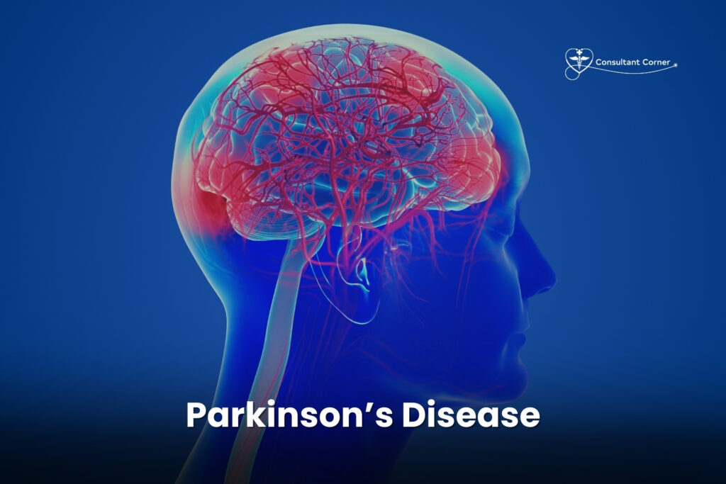
Parkinson’s Disease & Movement Disorders: 7 Warning Signs That Need Expert Evaluation Parkinson’s Disease rarely arrives all at once. For most people, it starts with one small, easy-to-dismiss change — a finger that trembles at rest, a shirt button that suddenly takes twice as long to fasten, handwriting that keeps getting smaller, or a walk that has quietly lost its usual rhythm. Because these shifts happen gradually, many people wait months or even years before asking a doctor about them. That delay matters. Parkinson’s Disease and other movement disorders respond best to treatment when they’re caught early, which is why an expert neurological evaluation — not guesswork — should be the first step. At Consultant Corner, we see patients every week who are trying to figure out whether what they’re experiencing is normal aging, stress, a side effect of medication, or something more. This guide walks through what Parkinson’s Disease actually is, the early warning signs your body may already be giving you, what else can mimic it, and what a real evaluation and treatment plan looks like. Is Tremor, Slowness, or Stiffness Starting to Affect Your Life? If you’ve noticed a tremor, a slower pace, new stiffness, or changes in your handwriting over the last several months, you’re not alone — and you’re not imagining it. Parkinson’s Disease and related movement disorders affect nearly one million people in the United States, and that number is expected to keep climbing as the population ages. These symptoms can chip away at independence, from struggling to open jars to feeling unsteady on stairs, and they tend to progress faster without intervention. A movement-disorder specialist can help slow that progression, manage symptoms, and keep you active and independent for longer. ⚠️ Early Warning Signs You Shouldn’t Ignore Parkinson’s Disease symptoms fall into two broad categories, and the non-motor ones are the group most people overlook. 🟣 Motor Symptoms Tremor in the hands or fingers, especially at rest Slowed movement (bradykinesia) or difficulty initiating tasks Stiffness in the arms, legs, or neck Balance problems or frequent falls Shuffling gait, reduced arm swing, or episodes of “freezing” while walking 🔵 Non-Motor Symptoms (Often Overlooked) Sleep problems, including acting out dreams physically Anxiety, depression, or loss of motivation Constipation or bladder changes Lightheadedness when standing up Loss of sense of smell Memory or concentration difficulties Research shows several of these non-motor symptoms — particularly REM sleep behavior disorder and loss of smell — can appear years before a formal diagnosis of Parkinson’s Disease is made, according to the National Institute of Neurological Disorders and Stroke. That’s exactly why early evaluation matters: the earlier a specialist can look, the earlier a plan can start. How Common Is This Condition? Movement disorders are far more common than most people assume. According to the Parkinson’s Foundation, nearly 90,000 people in the U.S. are diagnosed each year, and prevalence rises sharply with age. Men are somewhat more likely to be diagnosed than women, and while the condition is best known for its motor symptoms, researchers now understand that the underlying process affecting the brain often begins years, sometimes a decade or more, before a tremor ever becomes noticeable. That long runway is part of why awareness of subtle early signs — rather than waiting for a dramatic symptom — has become such a focus in modern neurology. 🔍 What Causes Parkinson’s Disease? Parkinson’s Disease develops when dopamine-producing neurons in the brain gradually decline, disrupting the signals that control smooth, coordinated movement. But here’s the part patients are often surprised by: not every tremor or every slow-moving day means Parkinson’s. Several other conditions can look remarkably similar on the surface, which is exactly why a proper neurological workup — not a quick guess — is so important. At Consultant Corner, we evaluate the full range of movement disorders, including: Parkinson’s Disease Essential tremor Drug-induced movement disorders Atypical Parkinsonism (PSP, MSA, CBD) Gait disorders Restless leg syndrome Post-stroke movement disorders You don’t have to self-diagnose from a symptom checklist online — that’s our job, and getting it right the first time changes the entire treatment path. How a Neurological Evaluation Actually Works A first visit for suspected movement symptoms typically isn’t a single quick test — it’s a process. A specialist will usually start by asking detailed questions about when symptoms began, whether they started on one side of the body or both, and whether they’ve changed with certain medications. From there, the exam looks at arm swing while walking, the speed and size of repetitive hand movements, muscle tone when the arm is moved passively, and how a resting tremor behaves compared to one that appears during voluntary movement. Balance is tested with a simple pull test, and handwriting samples can reveal micrographia — a telltale shrinking of letters that’s common in early Parkinson’s Disease. Blood work is often ordered to rule out thyroid dysfunction, vitamin deficiencies, or metabolic causes that can mimic parkinsonian symptoms. Imaging, such as an MRI, is generally used to exclude stroke, tumors, or structural abnormalities rather than to confirm Parkinson’s Disease directly, since the condition is primarily diagnosed clinically. The Stages of Parkinson’s Disease Understanding where symptoms fall on the disease timeline helps guide treatment decisions. Clinicians often reference a five-stage scale: Stage 1: Mild symptoms, usually on one side of the body only, with little or no impact on daily life. Stage 2: Symptoms affect both sides of the body; daily tasks take longer but independence is preserved. Stage 3: Balance becomes noticeably impaired and falls become more likely, though the person is still largely independent. Stage 4: Symptoms are severe enough that assistance is needed for some daily activities. Stage 5: Advanced stiffness or rigidity may make walking impossible without help; full-time care is often required. Most people spend years in the earlier stages, especially when treatment begins promptly — another reason early evaluation is worth prioritizing rather than postponing. Living With Parkinson’s Disease: What Helps Day to Day Medication is only part
Multiple Sclerosis: 12 Warning Signs and Expert Neurology
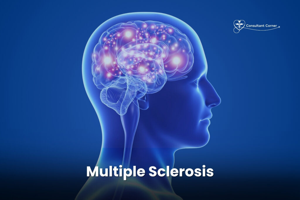
Multiple Sclerosis: 12 Warning Signs and Expert Neurology Evaluation Multiple Sclerosis can cause fatigue, weakness, vision changes, numbness, or balance problems that come and go — or slowly worsen over time. Whether you have just been diagnosed, you are worried about new symptoms, or you are searching for better long-term control, an early evaluation by a neurology specialist can make a meaningful difference in your quality of life. At Consultant Corner, our team helps patients understand what is happening in their bodies, stabilize their symptoms, and take back control of daily living. This guide walks through what Multiple Sclerosis actually is, the 12 warning signs you should never ignore, how the condition is diagnosed, what treatment looks like today, and how our Bloomington and El Paso neurology teams support patients through every stage of care. What Is Multiple Sclerosis? Multiple Sclerosis, often shortened to MS, is a chronic autoimmune condition in which the body’s immune system mistakenly attacks myelin — the protective coating that surrounds nerve fibers in the brain, spinal cord, and optic nerves. When myelin is damaged, electrical signals traveling along the nerves slow down or become distorted. The result is a wide and often unpredictable range of neurological symptoms. Because MS affects the central nervous system, its symptoms can appear almost anywhere in the body. No two patients experience the disease the same way, which is part of why an accurate diagnosis from an experienced neurologist is so important. Some people have mild, occasional flare-ups, while others face a more steadily progressive course. According to the National Multiple Sclerosis Society, nearly one million people are living with MS in the United States alone, and most are diagnosed between the ages of 20 and 50. 12 Early Warning Signs of Multiple Sclerosis You Shouldn’t Ignore MS affects everyone differently, but certain symptom patterns show up again and again. If you notice several of the signs below — especially if they come in episodes or gradually worsen — it may be time to schedule a neurology evaluation. Sensory Changes Numbness or tingling in the arms, legs, or face Electric-shock sensations that run down the spine or limbs, often triggered by bending the neck Unexplained weakness in an arm or leg Vision Problems Blurry or double vision Pain when moving the eyes Partial or temporary vision loss in one eye Mobility Challenges Balance problems or a feeling of unsteadiness Trouble walking or a change in gait Leg stiffness, spasms, or heaviness Cognitive and Energy Symptoms Brain fog, slower thinking, or memory lapses Fatigue that does not improve with rest Bladder and Bowel Changes Urinary urgency, frequency, or incontinence, along with constipation If these symptoms appear in flares that come and go, or if they are gradually getting worse, it’s worth discussing them with a specialist. Early evaluation of MS symptoms gives you and your care team more options and more time to protect long-term brain health. Understanding the Different Types of Multiple Sclerosis MS is not a single, uniform disease — it has several recognized subtypes, and the course it takes can differ significantly from one patient to the next. You do not need to know which type you have before seeking care; identifying the subtype is part of what a specialist evaluation is for. MS Subtype How It Typically Presents General Course Relapsing-Remitting MS (RRMS) Clear flare-ups (relapses) followed by periods of partial or full recovery Most common form at diagnosis, roughly 85% of cases Secondary Progressive MS (SPMS) Begins as RRMS, then gradually shifts to a steadier decline with fewer distinct relapses Often develops years after an initial RRMS diagnosis Primary Progressive MS (PPMS) Steady worsening of symptoms from the start, without early relapses and remissions Roughly 10–15% of cases Clinically Isolated Syndrome (CIS) A single episode of neurological symptoms lasting 24 hours or more May or may not progress to a definite MS diagnosis Why the Subtype Matters Knowing which form of MS a patient has helps guide treatment decisions, from choosing an appropriate Disease-Modifying Therapy (DMT) to setting realistic expectations for symptom management and long-term monitoring. What Causes Multiple Sclerosis? The exact cause of MS is still being studied, but researchers believe it results from a combination of factors rather than a single trigger. Current research points to: Immune system dysfunction — the immune system mistakenly identifies myelin as a threat and attacks it Genetic susceptibility — having a close relative with MS modestly increases risk, although MS is not directly inherited Environmental factors — including low vitamin D levels, certain viral infections such as Epstein-Barr virus, and smoking Geography — MS is more common in regions farther from the equator, which has led researchers to study the role of sunlight and vitamin D None of these factors alone explains why one person develops MS and another does not, which is why ongoing research and individualized medical evaluation remain essential. Multiple Sclerosis Statistics Worth Knowing Understanding how common MS is — and who it tends to affect — can help put a new diagnosis into perspective: Nearly one million Americans are currently living with MS, according to the National Multiple Sclerosis Society. MS is roughly two to three times more common in women than in men. Most people receive a diagnosis between the ages of 20 and 50, though pediatric and late-onset cases do occur. Relapsing-Remitting MS accounts for the large majority of diagnoses at the time of initial evaluation. With modern Disease-Modifying Therapies, many patients today experience fewer relapses and slower disease progression than patients diagnosed a generation ago. These numbers are encouraging, but they also underscore why a specialist evaluation matters so much. Statistics describe populations — your own MS journey depends on your specific symptoms, imaging findings, and how quickly you start an individualized treatment plan. How Is Multiple Sclerosis Diagnosed? There is no single test that confirms MS. Instead, diagnosis relies on a combination of clinical evaluation and imaging. At Consultant Corner, our diagnostic process typically includes: Neurological Examination A thorough
What Causes Ischemic Stroke? 8 Major Risk Factors Explained
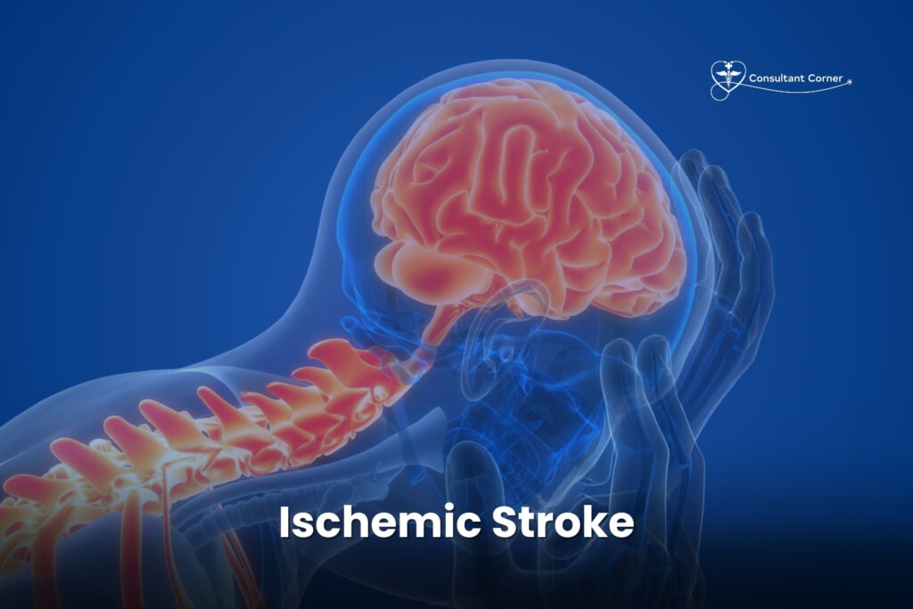
What Causes Ischemic Stroke? 8 Major Risk Factors Explained Ischemic stroke is the medical term for the most common type of stroke, and understanding what triggers one is the first step toward preventing it. An ischemic stroke happens when a blood vessel carrying oxygen-rich blood to the brain becomes blocked, usually by a blood clot or a buildup of fatty plaque. Without that blood supply, brain cells begin to die within minutes — which is why doctors often call stroke a “brain attack.” If you or someone you love has recently experienced stroke-like symptoms, or you simply want to understand your own risk, this guide walks through exactly what causes an ischemic stroke, who is most vulnerable, and what you can do about it. At Consultant Corner, our neurology team sees patients across Bloomington, Illinois and El Paso, Texas who are trying to make sense of a stroke diagnosis or a frightening set of symptoms. This article breaks the topic down in plain language so you can recognize the warning signs, understand your personal risk factors, and know when it’s time to seek expert care. What Is an Ischemic Stroke? An ischemic stroke accounts for roughly 87% of all strokes, making it far more common than the other major type, hemorrhagic stroke, which occurs when a blood vessel ruptures rather than becomes blocked. During an ischemic stroke, a clot or fatty deposit obstructs an artery feeding the brain. Brain tissue downstream of that blockage is starved of oxygen and nutrients, and without rapid treatment, that tissue can be permanently damaged. Every 40 seconds, someone in the United States has a stroke, and more than 795,000 people experience a new or recurrent stroke each year, according to the CDC’s Stroke Facts. The vast majority of those events are ischemic in nature, which is exactly why understanding the mechanism behind them matters so much for prevention. There are two main categories of ischemic stroke: Cerebral Thrombosis This occurs when a blood clot (thrombus) forms directly inside an artery that has already been narrowed by plaque buildup, a condition called atherosclerosis. The clot grows large enough to block blood flow at that exact location in the brain. Cerebral Embolism This happens when a clot forms somewhere else in the body — often in the heart — and then travels through the bloodstream until it lodges in a smaller brain artery. Irregular heart rhythms, especially atrial fibrillation, are one of the most common sources of these traveling clots. A brief version of an ischemic stroke, called a transient ischemic attack (TIA) or “mini-stroke,” happens when the blockage clears on its own within minutes. A TIA should never be dismissed — it is frequently the earliest warning sign that a larger, more damaging stroke may follow. If you’ve experienced brief symptoms that resolved on their own, our related article on TIA (Mini-Stroke) explains why prompt evaluation still matters even after symptoms disappear. What Causes Ischemic Stroke? The Root Mechanism At its core, every ischemic stroke traces back to something interrupting blood flow to the brain. The most frequent underlying cause is atherosclerosis — the slow, often silent buildup of cholesterol and fatty plaque inside artery walls. Over years, this plaque can narrow an artery enough to choke off blood flow on its own, or it can rupture and trigger a clot that finishes the job. Other, less common causes include: Blood clotting disorders that make the blood more prone to forming clots Inflammation of blood vessels (vasculitis) Dissection, or a small tear, in an artery wall supplying the brain Severe dehydration or blood-thickening conditions Certain infections that affect blood vessel health Because the causes of an ischemic stroke are so closely tied to the health of your blood vessels and heart, the risk factors below overlap heavily with cardiovascular disease. In fact, research on population-attributable risk has found that a relatively short list of well-known risk factors — hypertension, atrial fibrillation, physical inactivity, low HDL cholesterol, smoking, diabetes, and a few others — together account for nearly all of the risk of having an ischemic stroke in the first place. 8 Major Risk Factors for Ischemic Stroke Some risk factors for ischemic stroke can be modified through lifestyle changes and medical treatment, while others cannot. The table below summarizes the most significant ones. Risk Factor Type Why It Matters High blood pressure Modifiable The single leading cause of stroke; damages and weakens artery walls over time Atrial fibrillation / heart disease Modifiable Allows clots to form in the heart and travel to the brain High cholesterol Modifiable Drives plaque buildup (atherosclerosis) inside brain-supplying arteries Diabetes Modifiable Damages blood vessels and accelerates plaque formation Smoking or vaping Modifiable Thickens blood and damages vessel linings Obesity / sedentary lifestyle Modifiable Raises blood pressure, cholesterol, and diabetes risk simultaneously Prior stroke or TIA Non-modifiable (but manageable) Significantly raises the chance of a repeat, often more severe, event Age, family history, and sickle cell disease Non-modifiable Genetics and age naturally increase vulnerability 1. High Blood Pressure High blood pressure is considered the leading cause of stroke. Roughly three-quarters of people who have a stroke have hypertension, and it is often called the single most potent modifiable risk factor for both ischemic stroke and hemorrhagic stroke. Over time, uncontrolled blood pressure weakens and damages artery walls throughout the body, including the vessels that feed the brain, making them far more likely to narrow, tear, or clot. 2. Atrial Fibrillation and Heart Disease An irregular heartbeat allows blood to pool and clot inside the heart’s chambers. Those clots can break free and travel directly to the brain, causing an embolic ischemic stroke. People with atrial fibrillation face a substantially higher risk of stroke than the general population, which is why heart-rhythm monitoring is such a routine part of a neurologic workup after any stroke-like event. 3. High Cholesterol Excess LDL (“bad”) cholesterol builds up as plaque along artery walls, gradually narrowing the vessels that supply the brain and setting the
What Causes Intracerebral Hemorrhage? 7 Key Causes You Need to Know
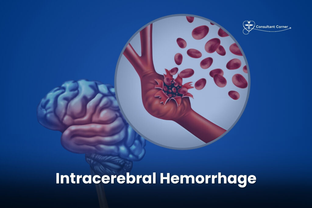
What Causes Intracerebral Hemorrhage? 7 Key Causes You Need to Know Intracerebral hemorrhage is one of the most dangerous types of stroke a person can experience, and understanding what triggers it can mean the difference between prevention and a life-altering emergency. Intracerebral hemorrhage occurs when a blood vessel inside the brain ruptures and spills blood directly into the surrounding brain tissue, damaging cells and raising pressure inside the skull within minutes. Unlike an ischemic stroke, which develops when a blood clot blocks blood flow, an intracerebral hemorrhage is a bleeding stroke — and it tends to strike suddenly, without any warning symptoms beforehand. At Consultant Corner, our neurology team sees firsthand how intracerebral hemorrhage changes lives overnight. This guide breaks down exactly what causes intracerebral hemorrhage, who is most at risk, how doctors diagnose and treat it, and what recovery really looks like — so you and your family know what to watch for and when to act. What Is Intracerebral Hemorrhage? Intracerebral hemorrhage (often shortened to ICH) is bleeding that happens within the brain’s own tissue, known as the parenchyma. It’s different from a subdural or epidural hemorrhage, which occur between the brain and skull, and different from a subarachnoid hemorrhage, which involves the space around the brain. When a vessel bursts inside the brain itself, blood pools into a mass called a hematoma. That hematoma compresses nearby brain tissue, cuts off oxygen supply, and can rapidly raise intracranial pressure — a combination that makes intracerebral hemorrhage a true medical emergency. This condition accounts for roughly 10-15% of all strokes in the United States, but it causes a disproportionate share of stroke-related deaths and long-term disability. Nearly half of all patients do not survive the first year, and among those who do, fewer than one in five regain full independence within six months. Outcomes depend heavily on how large the bleed is, exactly where it’s located, whether it extends into the brain’s fluid-filled ventricles, and — critically — how quickly treatment begins. Unlike ischemic strokes, which are sometimes preceded by a “mini-stroke” or transient ischemic attack that offers a warning, this type of brain bleed typically appears with no notice at all. One moment a person feels fine; the next, they’re experiencing a sudden headache, weakness, or confusion. That unpredictability is exactly why understanding the underlying causes — and managing the risk factors within your control — matters so much. Prevention is, by far, the most effective treatment available. The Main Causes of Intracerebral Hemorrhage Doctors generally divide the causes of intracerebral hemorrhage into two categories: primary (spontaneous) hemorrhage, which stems from long-term damage to small blood vessels, and secondary hemorrhage, which results from an identifiable structural problem like a tumor or malformed vessel. Here are the seven causes we see most often. 1. Chronic High Blood Pressure (Hypertension) Uncontrolled hypertension is the single leading cause of intracerebral hemorrhage. Over years, high pressure weakens the walls of small, deep arteries in the brain — particularly in areas like the basal ganglia, thalamus, cerebellum, and brainstem. Eventually those fragile vessel walls can rupture. This is why blood pressure management is often the first thing your neurologist addresses after an intracerebral hemorrhage diagnosis, and why it remains the most preventable cause of this condition. 2. Cerebral Amyloid Angiopathy (CAA) In older adults, a condition called cerebral amyloid angiopathy is a leading cause of intracerebral hemorrhage, especially bleeds that occur near the surface of the brain (lobar hemorrhages). CAA happens when an abnormal protein called amyloid builds up in the walls of small and medium blood vessels, making them stiff, brittle, and prone to rupture. It becomes more common with age and can sometimes accompany Alzheimer’s-related changes in the brain. 3. Head Trauma A fall, car accident, sports injury, or blow to the head can tear blood vessels and directly cause a traumatic intracerebral hemorrhage. Trauma-related bleeding can appear immediately or develop over the following hours or days, which is why anyone who experiences a significant head injury should be monitored closely, even if they initially feel fine. 4. Aneurysms and Vascular Malformations A brain aneurysm is a weak, bulging spot in an artery wall. Similarly, an arteriovenous malformation (AVM) is a tangle of abnormal blood vessels that connects arteries and veins without the normal capillary network in between. Both conditions create structurally weak points that can rupture and bleed into brain tissue, and both are more frequently identified as the cause of intracerebral hemorrhage in younger patients who don’t have hypertension or CAA. 5. Blood Thinners and Clotting Disorders Medications such as warfarin, heparin, and newer anticoagulants (like apixaban or rivaroxaban) reduce the blood’s ability to clot, which is essential for preventing conditions like ischemic stroke and blood clots — but it also raises the risk of bleeding if a vessel is injured or ruptures. Bleeding disorders, liver disease, and low platelet counts can have the same effect, making any brain bleed harder to control once it starts. 6. Drug and Alcohol Use Stimulant drugs, particularly cocaine and methamphetamine, cause sudden spikes in blood pressure that can trigger an intracerebral hemorrhage even in people with no prior vascular disease. Heavy, chronic alcohol use is independently linked to a higher risk of intracerebral hemorrhage as well, both by raising blood pressure and by impairing normal blood clotting. 7. Brain Tumors and Hemorrhagic Conversion Certain brain tumors have a rich, fragile blood supply and can bleed spontaneously. Intracerebral hemorrhage can also occur as a complication of a prior ischemic stroke — a process called hemorrhagic conversion — when the damaged blood vessels in a stroke-affected area become leaky as blood flow is restored. Can This Type of Brain Bleed Be Prevented? Because the leading cause is chronic high blood pressure, prevention largely comes down to long-term cardiovascular health. Steps that meaningfully lower risk include: Keeping blood pressure consistently under control with medication, home monitoring, and regular checkups Avoiding recreational stimulant drugs, including cocaine and methamphetamine Limiting alcohol intake to moderate levels Quitting
Headache & Migraine – Expert Evaluation & Relief
Headache & Migraine – Expert Evaluation & Relief Headache & Migraine pain is one of the most common — and most dismissed — reasons people put off getting help. If you’ve been told your pounding head, throbbing temples, or daily tension is “just stress,” but the pain keeps coming back, it may be time for a real evaluation instead of another over-the-counter pill. Recurring head pain isn’t something you have to accept as normal, and in most cases, it isn’t something you have to live with forever either. At Consultant Corner (Neurological Care in Bloomington, IL), our neurology team evaluates every type of headache and migraine disorder, from occasional tension headaches to disabling chronic migraine. Below, we’ll walk through the warning signs, the most common headache types, why untreated head pain matters, and what a real Headache & Migraine evaluation actually looks like — plus where a same-week visit or a walk-in clinic partner can fit into your care plan. Image placeholder: Neurologist reviewing brain scan with patient — Headache & Migraine evaluation — Alt text: “Headache & Migraine evaluation with a Bloomington IL neurologist” Headaches Taking Over Your Life? We Can Help. Frequent headaches or migraines aren’t “just stress” — they can deeply affect your work, sleep, mood, and daily life. Whether the pain is dull, sharp, throbbing, or one-sided, you deserve answers and relief. At Consultant Corner, we evaluate all types of headaches and help you get back to feeling like yourself again. Head pain is deeply personal. Some people describe it as a tight band squeezing their skull. Others describe migraine as a pulsing, nauseating wave that makes light and sound unbearable. No matter how your Headache & Migraine symptoms show up, the goal of an evaluation is the same: find the underlying cause, not just mask the pain. ⚠️ When Headaches Become a Problem Everyone gets headaches from time to time. A late night, dehydration, or a stressful afternoon can trigger a mild one that fades on its own. But certain patterns suggest something deeper may be going on, and that’s when a proper Headache & Migraine evaluation becomes important. You may need a specialist if you experience: Pain that returns weekly or daily Throbbing headaches with nausea or light sensitivity Headaches that worsen with activity or stress Pressure or tightness around the forehead or temples Headaches that disrupt sleep or work Pain around one eye with tearing or redness Headaches after a head injury If any of these sound familiar, your headaches are trying to tell you something. If you’ve recently experienced a fall, sports injury, or car accident, it’s also worth reading our related article on What is a Head Injury?, since post-traumatic headaches often need a different treatment approach than everyday tension headaches. If headaches are affecting your quality of life — even a little — there is a reason. And there is treatment. 🔍 Common Headache Types We Treat Not all head pain is created equal, and treating a migraine the same way you’d treat a tension headache rarely works. Here’s a quick breakdown of the most common types we see in our neurology clinics. Tension Headaches A constant pressure or “tight band” around the head, often linked to stress, posture, or muscle tension. Tension headaches are the most common type overall and often respond well to lifestyle changes, posture correction, and stress management. Migraines Severe, pulsating pain often with nausea, vomiting, light sensitivity, sound sensitivity, or vision changes (aura). Migraines can last hours to days and significantly limit daily functioning. According to the American Migraine Foundation, migraine is a disabling neurological disease rather than simply “a bad headache,” which is why it needs its own diagnostic approach and treatment plan. If you’re trying to pin down what sets off your attacks, our related post on 11 Migraine Triggers That Could Be Causing Your Headaches digs deeper into the most common culprits. Cluster Headaches Intense burning or piercing pain around one eye, occurring in sudden, severe bursts. These headaches are rare but extremely painful, and they often come with tearing, redness, or a drooping eyelid on the affected side. Secondary Headaches Headaches caused by underlying problems such as sinus issues, high blood pressure, medication overuse, dehydration, or neurological disorders. These require identifying and treating the root cause, not just the pain itself. You don’t need to know which type you have — that’s exactly what a Headache & Migraine evaluation is for. Here’s a simple side-by-side comparison to help you recognize the pattern that matches your symptoms: Headache Type Typical Pain Description Common Triggers Usual Duration Tension Headache Dull, tight, band-like pressure Stress, poor posture, fatigue 30 minutes – several hours Migraine Throbbing, pulsating, often one-sided Hormones, light, certain foods, stress 4 hours – 3 days Cluster Headache Sharp, burning, around one eye Alcohol, strong smells, sleep changes 15 minutes – 3 hours (recurring) Secondary Headache Varies (dull, sharp, or pressure) Sinus issues, medication overuse, high blood pressure Varies with underlying cause ❗ Why You Shouldn’t Ignore Recurring Headaches Untreated headaches may be related to: Neurological issues Stress and sleep problems Vision strain High blood pressure Post-concussion effects Medication overuse Chronic migraine syndrome Early evaluation helps determine the root cause and prevents symptoms from becoming chronic. In some cases, recurring headaches can also be an early warning sign tied to vascular health. Our vascular neurology team, led by Saqib A. Chaudhry, MD — a Vascular Neurologist and Neuro-intensivist — routinely evaluates patients whose headache patterns overlap with other neurological risk factors, including those covered in our articles on What Causes Ischemic Stroke? and 12 Early Ischemic Stroke Symptoms That Could Save Your Life. Headaches can also be an early symptom worth monitoring in patients with a family history of cognitive decline. If memory concerns run in your family alongside chronic headaches, our pieces on Understanding Alzheimer’s Disease and 10 Early Alzheimer’s Disease Symptoms You Should Never Ignore may also be a helpful read. The bottom line: a Headache & Migraine pattern
What is a Head Injury?
What Is a Head Injury? 7 Warning Signs to Know A head injury is any trauma to the scalp, skull, or brain caused by a fall, a car accident, a sports collision, or any other blow to the head. It can range from a mild bump that leaves nothing more than a tender lump, to a serious traumatic brain injury that changes how a person thinks, moves, or feels for months or years afterward. The tricky part about a head injury is that it doesn’t always look serious right away. Someone can walk away from an accident feeling “fine,” only to develop a pounding headache, confusion, or nausea hours or even days later. At Consultant Corner, we evaluate patients after falls, car accidents, workplace injuries, and sports collisions every week. Our goal with this guide is simple: help you understand what a head injury actually is, walk through the 7 warning signs that matter most, and explain when it’s time to see a neurologist rather than waiting it out at home. What Is a Head Injury, Exactly? Medically speaking, a head injury refers to any damage to the scalp, skull, or brain tissue. Doctors generally sort this trauma into two broad categories: Closed injury – the skull remains intact, but the brain can still be shaken, bruised, or bleed internally from the force of the impact. Open (penetrating) injury – the skull is fractured or punctured, and an object or fragment enters the brain tissue directly. Within these categories, a head injury is further described by severity: Mild (often called a concussion) – brief confusion, headache, or dizziness with no lasting structural damage on imaging. Moderate – longer periods of confusion or unconsciousness, sometimes with visible findings on a CT or MRI scan. Severe – significant bleeding, swelling, or structural damage that requires immediate emergency care and often surgery. It’s worth repeating: a head injury doesn’t have to involve a visible wound, bruise, or loss of consciousness to be serious. Many patients who later turn out to have a concussion, or even a small brain bleed, never “blacked out” at all. According to the Mayo Clinic, even injuries described as “mild” are still serious and require prompt attention and an accurate diagnosis. Common Causes of a Head Injury A blow to the head can happen in almost any setting, but the most frequent causes we see include: Falls at home, on stairs, or on ice (especially common in older adults and young children) Motor vehicle and motorcycle accidents Sports and recreational injuries, particularly contact sports and cycling Workplace accidents, including falls from height or being struck by equipment Physical assaults or violence Understanding the cause matters clinically. A low-speed fall in an older adult on blood thinners, for example, carries a very different risk profile than a teenager who was momentarily dazed during a soccer game — and both deserve a proper evaluation for a possible head injury. The 7 Warning Signs of a Head Injury You Should Never Ignore Because the brain’s function can be disrupted even without a visible wound, it’s important to watch closely for symptoms in the hours and days after any blow to the head. Here are the seven warning signs our neurology team tells every patient to watch for: # Warning Sign What It Can Mean 1 Headache that won’t go away or keeps getting worse Can be an early sign of concussion or bleeding inside the skull 2 Dizziness, imbalance, or feeling “off” Often signals disruption in the brain pathways that control coordination 3 Nausea or vomiting A common indicator of increased pressure inside the brain 4 Trouble focusing, remembering, or processing information Many patients describe feeling “foggy” or unusually slow afterward 5 Sensitivity to light or blurry vision Can appear days later and suggests irritation of the brain after trauma 6 Mood changes, irritability, or unusual emotional swings Reflects how trauma can affect the brain’s emotional regulation centers 7 Sleep disturbances (sleeping far more or far less than usual) A frequently overlooked but common after-effect of head injury If any of these seven signs show up — or get worse instead of better — it’s safest to be evaluated by a specialist promptly rather than waiting to see if things improve on their own. Symptoms That Can Take Days to Appear One of the most misunderstood things about a head injury is the delay in symptoms. Swelling, small amounts of bleeding, and inflammation inside the skull can build gradually, which means someone can feel completely normal on day one and then develop a severe headache, confusion, or vision changes on day three or four. The CDC notes that a traumatic brain injury affects how the brain works and can present with symptoms that show up well after the initial event. This delayed pattern is exactly why close monitoring is recommended for at least the first 24 to 48 hours after any significant impact, and continued attention for up to two weeks in cases involving concussion. Why a Head Injury Should Always Be Taken Seriously Outcomes are unpredictable, and what looks like a simple bump can sometimes lead to more serious complications, including: Concussion and lingering post-concussion symptoms that affect concentration, mood, and sleep for weeks or months Internal bleeding, such as an epidural or subdural hematoma, which can put dangerous pressure on the brain Skull fractures, which may or may not be visible from the outside Persistent headaches, memory issues, mood changes, or sleep problems, sometimes called post-concussion syndrome The National Institute of Neurological Disorders and Stroke (NINDS) explains that initial damage from a head injury can cause secondary damage — changes that unfold over hours to days after the original event — which is part of why early evaluation matters so much for long-term recovery. Catching a head injury early can prevent dangerous complications and dramatically improve recovery outcomes, especially when bleeding or swelling is caught before it becomes an emergency. Children vs. Adults: Does Age Change the Risk? Every
Epilepsy & Seizure Disorders: 7 Warning Signs and Treatment Options
Epilepsy & Seizure Disorders: 7 Warning Signs and Treatment Options Epilepsy & Seizure Disorders affect millions of people across the United States, yet they remain some of the most misunderstood conditions in neurology. Whether it causes brief “blank” staring moments, sudden jerking movements, or a full convulsive episode, a seizure can be frightening for the person experiencing it and for everyone watching. If you or a loved one has had even one unexplained seizure, getting evaluated quickly by a neurology specialist is one of the most important steps you can take for long-term brain health. At Consultant Corner, we specialize in diagnosing the underlying cause of seizures, building personalized treatment plans, and helping patients regain safety, confidence, and control over their daily lives. This guide walks through everything you need to know about Epilepsy & Seizure Disorders — from early warning signs to advanced treatment options — so you know exactly what to expect and when to seek help. Experiencing Seizures or Sudden Episodes? We Can Help. Epilepsy & Seizure Disorders can be unpredictable and disruptive to everyday life. A seizure doesn’t always look like the dramatic, full-body convulsion many people picture. Sometimes it’s a brief moment of confusion, a strange sensation, or a lapse in awareness that lasts only a few seconds. Because seizures present so differently from person to person, many go unrecognized for months or even years before a diagnosis is made. That’s why early evaluation matters. The sooner a seizure disorder is identified, the sooner a treatment plan can be put in place to reduce the frequency and severity of future episodes. 7 Warning Signs You May Need an Evaluation for Epilepsy & Seizure Disorders Seizures can look very different from person to person. You may need an evaluation if you’ve experienced any of the following: Sudden “blank staring” or unresponsive moments Repetitive movements, such as lip smacking or picking at clothing Jerking or shaking of the arms or legs Loss of consciousness or blackout spells Strange sensations, including unusual smells, tastes, tingling, or déjà vu episodes Sudden confusion or disorientation without an obvious cause Unexplained falls or collapses Even a single episode can be a sign of an underlying neurological condition that deserves attention. Ignoring these signs, or assuming they will resolve on their own, can delay a diagnosis and increase the risk of injury. What Is Epilepsy? Understanding Epilepsy & Seizure Disorders Epilepsy is a chronic neurological condition in which the brain produces sudden bursts of abnormal electrical activity, causing recurrent and unprovoked seizures. According to the Centers for Disease Control and Prevention, millions of American adults live with active epilepsy, making it one of the most common neurological conditions in the country. However, not every seizure means a person has epilepsy. A thorough evaluation is necessary to distinguish between: Epileptic seizures caused by abnormal brain electrical activity Non-epileptic episodes with a psychological or stress-related origin Fainting spells (syncope) that can resemble seizure activity Metabolic or medication-related spells, including low blood sugar or drug interactions Sleep-related seizure activity that occurs only during certain sleep stages You don’t need to know which category applies to you before scheduling an appointment — identifying the correct cause is exactly what our neurology team is trained to do. Types of Seizures We Diagnose and Treat Understanding the type of seizure a patient is experiencing is one of the most important steps in building an effective treatment plan for Epilepsy & Seizure Disorders. Below is a breakdown of the most common categories our specialists evaluate. Seizure Type Common Signs Awareness Level Focal Seizures Tingling, jerking, sudden emotions, déjà vu sensations May or may not involve loss of awareness Tonic-Clonic (Grand Mal) Stiffening of the body followed by rhythmic jerking, collapse Loss of consciousness Absence (Petit Mal) Brief staring spells, sometimes with eye fluttering Brief loss of awareness Atonic (“Drop Attacks”) Sudden loss of muscle tone, causing a fall Variable Myoclonic Quick, sudden body jerks or muscle twitches Usually retained Tonic or Clonic Only Stiffening or rhythmic jerking without a full convulsive pattern Variable Knowing the specific seizure type helps our specialists choose the medication, dosage, and lifestyle recommendations that are most likely to be effective for each individual patient. Common Causes and Risk Factors for Epilepsy & Seizure Disorders Understanding what may be behind a seizure disorder is a key part of building an effective treatment plan. Epilepsy & Seizure Disorders can develop for many different reasons, and in some cases, more than one factor is involved. Common causes and risk factors our specialists evaluate for include: Genetics — Some forms of epilepsy run in families or are linked to specific inherited gene mutations. Head injury — Traumatic brain injury from a fall, car accident, or sports injury can trigger seizures, sometimes years after the initial injury. Stroke — A prior ischemic or hemorrhagic stroke can leave scar tissue that disrupts normal electrical activity in the brain. Brain tumors or structural abnormalities — Growths or malformations in brain tissue can serve as a seizure focus. Infections — Conditions such as meningitis, encephalitis, or certain parasitic infections can damage brain tissue and increase seizure risk. Developmental conditions — Some children are born with brain differences that make them more prone to seizures. Metabolic or chemical imbalances — Severe changes in blood sugar, sodium, or other electrolytes can provoke seizure activity. Prenatal or birth-related complications — Oxygen deprivation or infection during pregnancy or delivery can be a contributing factor later in life. In roughly half of all cases, no clear cause is ever identified. This is known as idiopathic epilepsy, and it does not mean the condition is any less treatable — it simply means the underlying trigger cannot be pinpointed through current diagnostic tools. How We Diagnose Epilepsy & Seizure Disorders Getting an accurate diagnosis is the foundation of effective treatment. When you come to Consultant Corner for an evaluation, our process typically includes: A detailed intake conversation. We ask about what the episode looked like, how long it
Alzheimer’s Disease & Memory Loss – Early Action Can Change Everything
Alzheimer’s Disease & Memory Loss – Early Action Can Change Everything Alzheimer’s Disease rarely arrives with a single dramatic moment. For most families, it shows up in small pieces first — a missed bill payment, a story repeated twice in one phone call, a familiar drive that suddenly feels confusing. Because these moments look so much like ordinary aging, they’re easy to explain away, and that’s exactly the problem. The earlier Alzheimer’s Disease is recognized and evaluated, the more options a patient and their family have — for treatment, for planning, and for protecting independence as long as possible. At Consultant Corner, our neurology team meets with patients and caregivers every week who are trying to answer one question: is this normal aging, or is this something more? This guide walks through the warning signs worth paying attention to, the many conditions that can be mistaken for Alzheimer’s Disease, and — most importantly — the concrete steps that early action makes possible. Is Forgetfulness Starting to Impact Daily Life? Memory loss can be overwhelming, especially once it starts affecting conversations, appointments, familiar places, or personal routines. What feels like “just getting older” may actually be an early sign of Alzheimer’s Disease, Mild Cognitive Impairment (MCI), or another neurological condition entirely. The distinction matters. Occasionally misplacing your keys is normal. Finding them in the refrigerator, with no memory of putting them there, and then suspecting a family member moved them, is a different pattern. Alzheimer’s Disease tends to be progressive and disruptive — it interferes with the things a person has always done automatically, not just the things that were hard to begin with. Early evaluation can slow decline, protect independence, and give families clarity instead of guesswork. That clarity is often the first thing that brings relief to a worried spouse or adult child, even before treatment begins. Red Flags of Alzheimer’s Disease You Should Never Ignore If you notice any of the patterns below — especially more than one at the same time — a neurological evaluation is worth scheduling sooner rather than later. Memory Changes Forgetting recent events or entire conversations, rather than just a name or a date, is one of the clearest early indicators. So is misplacing items in unusual places and being unable to retrace your steps to find them. Thinking and Communication Problems Trouble finding the right word mid-sentence, repeating the same question multiple times in one visit, or losing the thread of a conversation are all signs worth noting. This goes beyond the occasional “it’s on the tip of my tongue” moment everyone experiences from time to time. Daily Function Decline Struggling to manage bills, medications, or appointments that used to be routine — or getting lost in a neighborhood the person has lived in for years — points toward a functional decline rather than simple forgetfulness. Behavioral or Mood Changes Irritability, anxiety, depression, or withdrawal from social activities can accompany early Alzheimer’s Disease. So can new suspicion, paranoia, or decisions that feel out of character, such as falling for a scam or giving away money unexpectedly. These warning signs often appear months, or even years, before a formal diagnosis is made. Recognizing the pattern early — rather than waiting for a crisis — is what gives a family the most room to plan. For a symptom-by-symptom breakdown, see our companion article, 10 Early Alzheimer’s Disease Symptoms You Should Never Ignore. Memory Loss Isn’t Always Alzheimer’s Disease One of the most reassuring things families learn during an evaluation is that memory loss has many possible causes, and Alzheimer’s Disease is only one of them. Part of a neurologist’s job is separating the treatable causes from the progressive ones, rather than assuming the worst from the start. Possible Cause Is It Treatable? What It Often Looks Like Alzheimer’s Disease Not reversible, but manageable Gradual, progressive memory and thinking decline Mild Cognitive Impairment (MCI) Sometimes stable, sometimes progresses Noticeable changes that don’t yet interfere with daily life Vitamin B12 or folate deficiency Yes, highly treatable Fatigue, confusion, memory lapses Thyroid disorders Yes, treatable with medication Brain fog, mood changes, slowed thinking Depression and anxiety Yes, treatable Poor concentration, apathy, forgetfulness Sleep disorders (including apnea) Yes, often treatable Daytime confusion, poor short-term memory Medication side effects Yes, adjustable Sudden confusion after a new prescription Mini-strokes (TIAs) or vascular dementia Requires vascular management Step-wise decline, sometimes with physical symptoms Past concussion or head injury Often improves with rehab and time Memory and concentration problems after trauma Because so many of these causes overlap in how they present, guessing at the reason from home rarely gives a clear answer. A structured evaluation is what actually uncovers the cause — and finding it early makes treatment more effective, whatever the underlying condition turns out to be. If a past concussion is part of the picture, our guide What is a Head Injury? is worth reading alongside this one. Why Early Diagnosis of Alzheimer’s Disease Matters Catching Alzheimer’s Disease early doesn’t change the underlying diagnosis, but it changes almost everything about what happens next. Early action generally means: Better symptom control through timely treatment A slower rate of cognitive decline Safer daily functioning, with fewer accidents or safety risks More time to plan legal, financial, and care decisions while the patient can still participate Access to more effective medication options Stronger support systems for caregivers and family members Waiting, on the other hand, tends to mean a faster decline and fewer treatment paths by the time care finally begins. If any of the red flags above sound familiar, it’s worth acting on that instinct now rather than waiting for a more obvious crisis. Who Is Most at Risk for Alzheimer’s Disease? Researchers don’t yet fully understand why some people develop this condition and others don’t, but several risk factors are well established. None of them guarantee a diagnosis on their own — they simply help patients and families understand where extra vigilance is worthwhile. Age The single biggest known
Types of Headaches Explained: Causes, Symptoms, and Duration
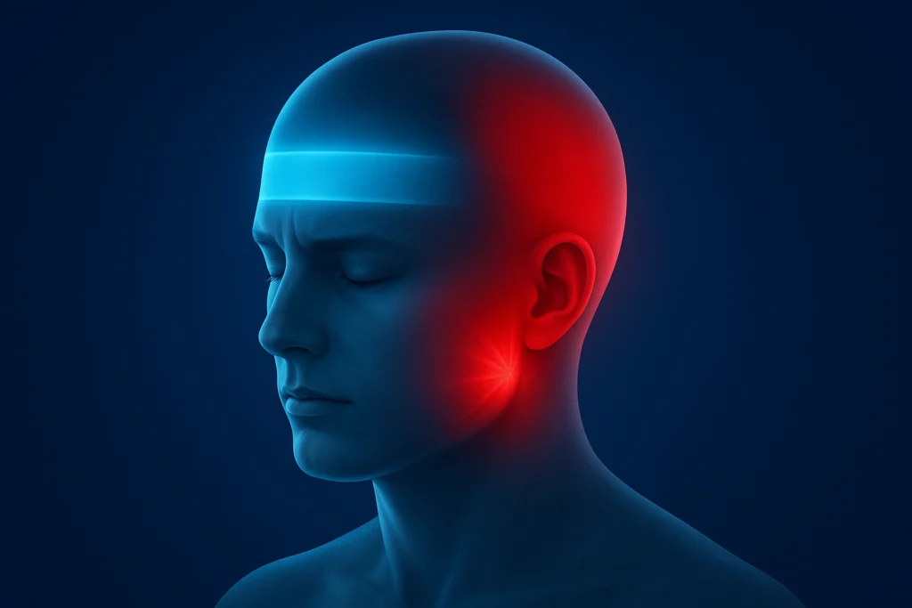
Types of Headaches Explained: Causes, Symptoms, and Duration Understanding the different types of headaches can help you identify what you’re experiencing and when to seek medical help. Headaches are broadly classified into primary headaches (not caused by another condition) and secondary headaches (caused by underlying health issues). Primary Headaches These occur independently and are not a symptom of another disease. 1. Tension Headaches Most common headache type Feels like a tight band or pressure around the head Pain level: mild to moderate Common triggers: stress, poor posture, muscle tension Duration: Minutes to several days 2. Migraine Headaches Severe, often one-sided pain May include nausea, vomiting, sensitivity to light or sound Can be preceded by an aura (vision changes, tingling sensations) Duration: Hours to days Migraines may significantly impact daily life and often require treatment. 3. Cluster Headaches Extremely painful, stabbing headache on one side, usually around the eye Occur in “clusters” – multiple attacks per day over weeks or months May cause watery eyes, nasal congestion, or eyelid drooping Duration: 15 minutes to 3 hours 4. Other Primary Headaches Triggered by specific actions such as coughing, exercise, or sleep (hypnic headaches) Symptoms and duration vary depending on the trigger Secondary Headaches These are caused by another health issue, such as: Infections Head or neck injuries Medication side effects Underlying medical conditions Summary Table: Common Primary Headaches Headache Type Description Common Symptoms Duration Tension Band-like pressure Mild to moderate pain Minutes to days Migraine One-sided, severe Nausea, aura, light/sound sensitivity Hours to days Cluster Stabbing pain near one eye Watery eyes, nasal congestion 15 min – 3 hrs Other (cough, exercise, hypnic) Triggered by actions Varies Varies When to See a Doctor Seek immediate medical attention if you experience headaches: Become more frequent or severe Are accompanied by weakness, confusion, or vision loss Follow an injury or sudden health change About Our Telehealth Neurology Services: We are Neurological Care Specialists, dedicated to providing expert evaluation and treatment exclusively through telehealth services. Our care covers a wide range of neurological conditions, including: Head Injury Ischemic Stroke Headache / Migraine Alzheimer’s Disease Parkinson’s Disease Multiple Sclerosis Epilepsy and Seizures Intracerebral Hemorrhage If you are located in Illinois or nearby areas, you can access our specialized neurological care conveniently from your home. 📞 Book your telehealth consultation today and take the first step toward better neurological health.
What is a Lenticulostriate Stroke?
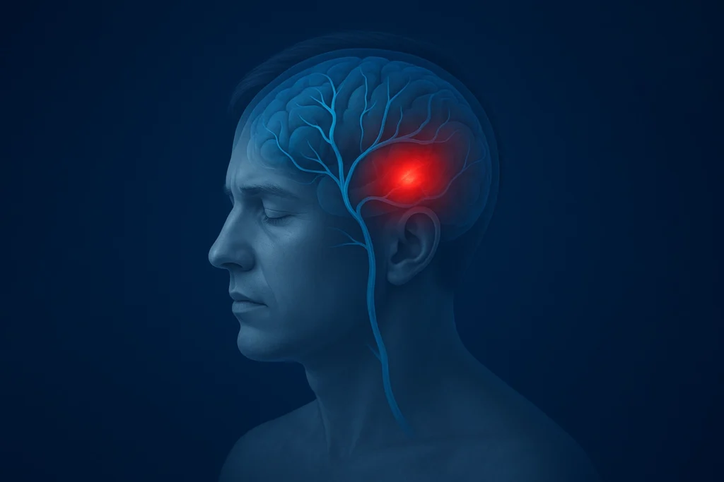
Sore Back and Dizziness | Telehealth Neurologist in Bloomington
Why Do I Have a Sore Back and Dizziness at the Same Time? Experiencing back soreness with dizziness can be unsettling. These symptoms often occur together due to problems in the spine, muscles, posture, or nervous system. In some cases, they may even signal an underlying neurological or vascular condition. Here’s a detailed look at what might be causing it — and when to seek medical help. Can Cervical Spine Problems Cause Dizziness? Yes. Conditions affecting the cervical spine (neck and upper back)—such as herniated discs, cervical spondylosis (arthritis of the neck), or muscle strain—can interfere with nerves and blood vessels responsible for balance. When these structures are compressed or irritated, it can lead to neck pain, upper back soreness, and dizziness. This condition is sometimes known as cervical vertigo, where neck-related issues trigger balance disturbances or lightheadedness. Can Poor Posture Lead to Dizziness and Back Pain? Absolutely. Spending long hours in poor posture—like slouching or forward head position—strains the muscles in your back and neck. This muscle tension can reduce blood flow to the brain and affect your vestibular system (which controls balance), resulting in both back soreness and dizziness. Modern lifestyles, which often involve computer work, phone use, or prolonged sitting, are common culprits behind this posture-related discomfort. Learn More: Can Bad Posture Cause Headaches Can Problems in the Spine or Neck Cause Dizziness? Yes — spinal and neck issues are among the most common causes of dizziness combined with back pain. When nerves in the neck or upper spine become compressed or irritated, they can interfere with the body’s balance centers in the brain. Conditions such as: Cervical herniated discs Cervical spondylosis (arthritis of the neck) Pinched nerves in the neck or upper back can lead to both neck soreness and dizziness, a condition often called cervicogenic dizziness or cervical vertigo. Can Poor Posture or Muscle Tension Cause Dizziness? Absolutely. Poor posture — especially forward head posture or slouching — puts excessive strain on the neck, shoulder, and upper back muscles. Over time, this muscle tension can compress nerves or reduce blood flow to the brain, resulting in lightheadedness, imbalance, and pain. This is common in people who sit for long hours at computers or look down at phones, and it may cause what’s known as postural or tension-related dizziness. Could Spinal Injuries or Trauma Be the Cause? Yes. Spinal cord injuries, whiplash, or severe back trauma can disrupt nerve signaling and balance mechanisms in the brainstem and inner ear. This can lead to symptoms such as: Back pain or stiffness Vertigo or spinning sensation Loss of coordination or unsteadiness Anyone who experiences dizziness after an accident or fall should seek medical care immediately to rule out spinal cord injury or concussion. How Do Muscle Tension and Nerve Irritation Contribute? When the upper back, neck, or shoulder muscles tighten excessively, they can press on nearby nerves and blood vessels, disrupting circulation and nerve signals. This tension may cause a combination of headaches, dizziness, muscle stiffness, and soreness. Chronic stress, lack of movement, or repetitive strain often worsen these symptoms. Can Whiplash or Spine Injuries Cause Dizziness? Yes. Traumatic injuries—such as whiplash from a car accident or sports injuries—can strain or damage neck muscles, ligaments, and spinal structures. These injuries may alter how your brain processes signals from your neck and inner ear, leading to pain, dizziness, and imbalance. Post-traumatic dizziness can persist for weeks or months if untreated. What Other Conditions Can Cause Dizziness with Back Pain? Some systemic or musculoskeletal conditions can also cause dizziness and back soreness, including: Osteoarthritis or sciatica Fibromyalgia Pregnancy (due to posture changes and circulation shifts) Dehydration or low blood sugar Vascular disorders affecting blood flow Since these symptoms can overlap with those of many conditions, accurate diagnosis is crucial. How Is Dizziness with Back Pain Treated? Treatment depends on the underlying cause but may include: Posture correction and ergonomic adjustments Physical therapy to strengthen neck and back muscles Stretching and muscle relaxation techniques Pain relievers or muscle relaxants (if prescribed) Vestibular rehabilitation for dizziness related to balance disorders If your dizziness is severe, persistent, or accompanied by numbness, vision problems, or difficulty walking, seek urgent medical attention. These could indicate a neurological or vascular emergency. What Symptoms Should I Watch For? Along with back pain, dizziness may appear as: Lightheadedness or faintness Feeling “off-balance” or unsteady Spinning sensation (vertigo) Blurred vision or disorientation Difficulty maintaining coordination If these symptoms are persistent, severe, or worsening, or if they occur with numbness, weakness, or vision changes, urgent medical evaluation is necessary. Such signs could indicate stroke, nerve compression, or vascular problems. When Should I See a Doctor? You should contact a healthcare provider or neurologist if you experience: Recurrent or worsening dizziness Severe neck or upper back pain Unsteadiness or balance loss Headaches, blurred vision, or confusion Recent trauma or whiplash Early evaluation can help identify treatable causes and prevent complications. How Can MyCornerConsultant Help? At MyCorner Consultant, we provide neurologist telehealth consultations for patients experiencing back pain, dizziness, and balance problems in Bloomington and all nearby cities. We specialize in identifying and treating neurological causes, including: Cervical vertigo and balance disorders Headache and migraine Spine-related nerve compression Stroke and vascular issues Traumatic head or neck injuries Through secure telehealth sessions, patients across Bloomington and all nearby cities can access expert neurological care without the need for in-person visits. Take the First Step Toward Relief If you’re struggling with persistent back soreness and dizziness, don’t ignore the signs — they could point to an underlying neurological or spinal condition. Our goal at MyCornerConsultant is to help you understand your symptoms, identify the cause, and create a clear recovery plan — all through accessible, patient-centered telehealth care. Schedule your online neurology consultation today and connect with a trusted Bloomington neurologist dedicated to your brain and spine health.
How Big of a Problem Are Headaches in the U.S
How Big of a Problem Are Headaches in the U.S.? Headaches are one of the most common—and often overlooked—public health issues in America. Affecting millions each year, they significantly impact quality of life, healthcare systems, and workplace productivity. From mild tension headaches to chronic migraines, these conditions remain underdiagnosed and undertreated, despite their growing burden on society. How Many Americans Experience Headaches? Headaches are nearly universal, but recent studies highlight the true scale: Around 78 million Americans experience migraines or severe headaches in their lifetime. Migraines alone affect about 12% of the population, or roughly 38 million people. Tension-type headaches occur in up to 80% of adults, typically linked to stress, fatigue, and posture. The National Headache Foundation reports that 1 in 6 Americans experiences headaches regularly. These figures confirm that headaches affect people of all ages, backgrounds, and lifestyles. How Much Do Headaches Cost the U.S. Economy? Headaches come with a high economic price tag—estimated at over $13 billion annually.This includes: Direct costs: physician visits, diagnostics, and medications Indirect costs: absenteeism, reduced performance, and lost productivity Every year, headaches are responsible for approximately 157 million lost workdays, making them a major driver of workplace inefficiency and absenteeism. How Do Headaches Strain the Healthcare System? Headaches rank among the top 15 reasons for emergency department visits in the United States.Millions of people seek urgent medical care for sudden or severe headaches, concerned about conditions such as stroke or meningitis. Chronic headache sufferers—particularly migraine patients—require frequent consultations, neurological care, and long-term management, driving up overall healthcare utilization and costs. Who Is Most Affected by Headaches? Headaches do not discriminate, but certain groups face a higher risk: Women are three times more likely than men to experience migraines, largely due to hormonal changes. Prevalence peaks between ages 30 and 50, coinciding with high work and family responsibilities. Children and older adults can also experience headaches, though diagnosis and treatment may differ by age. These demographic insights show the importance of tailored headache care across life stages. How Do Headaches Affect Daily Life and Well-Being? For many, headaches are more than just pain—they disrupt daily routines, relationships, and emotional health.Chronic or frequent headaches can lead to: Difficulty focusing or completing work Reduced participation in social or family activities Increased stress, anxiety, and sleep issues Lower overall quality of life Migraine sufferers, in particular, often experience significant disability during attacks, which may last from hours to days. Why Are Headaches a Major Public Health Concern? Headaches—especially migraines and tension-type headaches—affect tens of millions of Americans and cost billions in healthcare and lost productivity.They remain among the top neurological complaints and a major source of preventable disability. Increasing awareness, improving diagnosis, and promoting early management strategies are essential steps toward reducing this hidden but widespread public health burden. Expert Neurology Care — Available Across Bloomington and Beyond At My Corner Consultant, we bring compassionate, evidence-based neurology care directly to your screen — no long drives, no crowded waiting rooms, and no delays in getting answers. Our Bloomington-based neurologist offers trusted telehealth consultations for patients throughout Bloomington and all surrounding cities, ensuring everyone has access to specialized brain and nerve care, wherever they are. We diagnose and manage a wide range of neurological conditions, including: Head Injury & Concussion Management – Comprehensive evaluation and follow-up care. Ischemic Stroke & Post-Stroke Recovery – Expert guidance to prevent recurrence and improve function. Headache & Migraine Disorders – Tailored treatment plans to reduce frequency and intensity. Alzheimer’s Disease & Memory Disorders – Early detection, cognitive assessment, and ongoing support. Parkinson’s Disease & Movement Disorders – Medication management and quality-of-life care. Multiple Sclerosis (MS) – Symptom management and long-term care planning. Epilepsy & Seizures – Diagnosis, medication optimization, and lifestyle support. Intracerebral Hemorrhage & Brain Bleeds – Specialist consultation for recovery and prevention. Wherever you live in Bloomington or nearby areas, expert neurological care is just a video call away.Our goal is to make high-quality neurology accessible, personal, and convenient for every patient — because your brain health deserves nothing less. Telehealth Appointments Available in Bloomington and All Nearby Cities
Can Bad Posture Cause Headaches
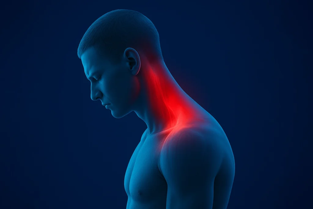
Can Bad Posture Cause Headaches? Yes, bad posture can cause headaches. Poor posture strains muscles in the neck, upper back, and shoulders, causing muscle tightness, spasms, and inflammation. This tension, especially around the base of the skull and neck, can irritate nerves and trigger cervicogenic headaches, which often start in the neck and radiate to the forehead, temples, or behind the eyes. Forward head posture, slumping, or hunching puts extra pressure on the cervical spine, forcing the neck muscles to overwork in support of the head’s weight. This leads to muscle fatigue and joint stress, which contribute to headaches. In addition, bad posture can restrict blood flow and compress nerves, worsening headache symptoms. Also, abnormal posture can alter breathing patterns, further exacerbating headaches. What Are the Symptoms of Bad Posture Headaches? Pain starting at the base of the skull, radiating to the temples, forehead, or behind the eyes Neck pain and stiffness worsen with head or neck movement Muscle tightness or spasms in the upper back and neck Possible tingling or numbness in the arms due to nerve compression Lightheadedness and difficulty concentrating in some cases Poor posture also reduces blood flow and may compress nerves, aggravating headache symptoms. Both slouching and overly rigid upright posture can contribute to creating muscle imbalances or improper breathing patterns. How Does Bad Posture Cause Headaches? Introducing structural imbalances such as forward head posture and a hyperkyphotic thoracic spine Causing muscle tension and spasms, particularly in the neck and upper back muscles Irritating nerve pathways that refer pain to the head, mimicking migraine or tension headaches How Can You Treat or Prevent Bad Posture Headaches? Treatment typically involves improving posture through: Physical therapy and ergonomic adjustments Stretching and strengthening exercises for neck and upper back muscles Correcting spinal alignment and maintaining neutral posture while sitting or standing Taking regular breaks from screens or desks to reduce strain How Poor Posture Contributes to Headaches? Poor posture is a significant contributor to headaches, with research highlighting its impact on neck pain and headache frequency. Forward head posture (FHP), a common form of bad posture, places excessive strain on the cervical spine and associated muscles, leading to cervicogenic headaches—a type of headache originating from the neck. Key Statistics and Research Findings Studies show a moderate negative correlation between forward head posture angle and neck pain intensity (correlation coefficient r = −0.536, p < 0.01). The prevalence of cervicogenic headache among healthcare workers, such as nurses, is approximately 27.1%, with neck disability reported in 31.4% of subjects in one study. Forward head posture increases the odds of headaches by about 4.9% for every 1 cm of head displacement from normal alignment. Around 45.4% of dentists experience cervicogenic headaches, linked to postural abnormalities like upper crossed syndrome. Overall, cervicogenic headaches affect between 0.4% and 42% of the general population, depending on study cohorts and diagnostic criteria. Mechanisms Linking Posture and Headache Development Poor posture-related headaches stem from muscle fatigue, nerve irritation, and joint stress in the cervical region. These headaches often present with neck pain, stiffness, and radiating pain from the neck to the head, and they can mimic tension or migraine headaches. How to Reduce Headaches Caused by Poor Posture Addressing posture through ergonomic interventions, physical therapy, and strengthening exercises for the neck and upper back has been shown to effectively reduce headache frequency and intensity associated with poor posture. Final Insights Bad posture is a well-documented yet under-recognized factor in headache prevalence. It’s strongly associated with neck pain and cervicogenic headaches in both general and occupational populations, emphasizing the need for postural awareness and preventive care. Telehealth Neurology Services in Illinois At MyConsultantCorner, we provide expert telehealth neurology services across Illinois, helping patients receive quality care without the need to travel. Whether you’re dealing with headaches, migraines, dizziness, neuropathy, or other neurological conditions, our board-certified neurologists are here to help through secure, convenient online consultations. We focus on understanding your symptoms, offering personalized treatment plans, and guiding you toward better brain and nerve health—all from the comfort of your home. Book your telehealth neurology appointment in Illinois today and get the professional care you deserve—anytime, anywhere.
What is a headache?
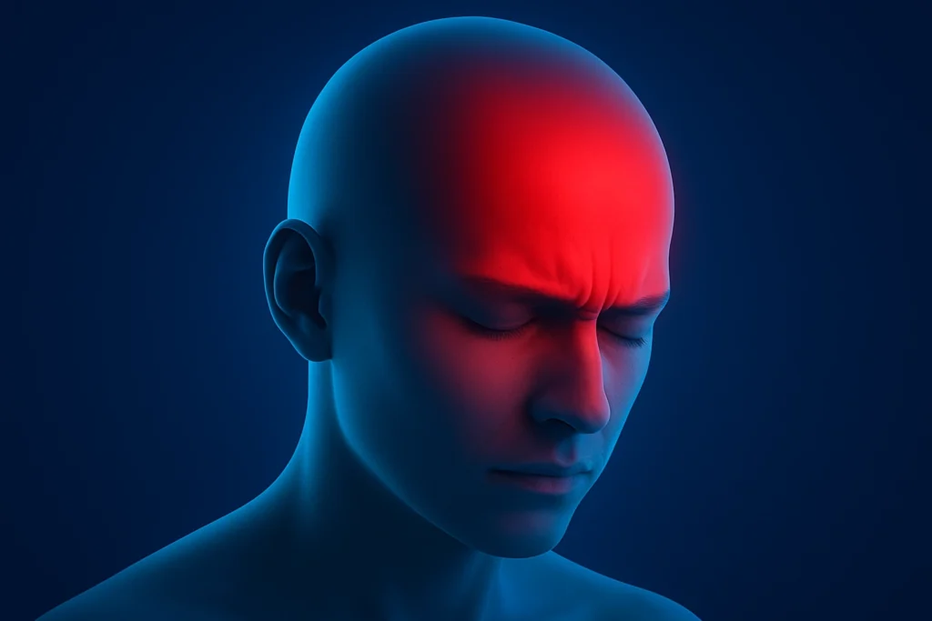
What is a headache? A headache is pain or discomfort that occurs in any part of the head, including the face, scalp, or upper neck. It is one of the most common physical complaints and can range from mild to severe in intensity. Headaches can be classified as primary, meaning they are not caused by another medical condition (such as tension headaches, migraines, or cluster headaches), or secondary, meaning they result from an underlying medical issue such as injury, infection, or other diseases. The pain in a headache comes from irritation or dysfunction of pain-sensitive structures around the skull, including muscles, nerves, blood vessels, and tissues. The brain itself does not have pain receptors. Headaches can feel like throbbing, pressure, tightness, or sharp pain, and symptoms may vary depending on the type of headache. Most headaches are not signs of serious illness, but severe, persistent, or unusual headaches warrant medical evaluation. What are the types of headaches? Headaches are generally classified into two main types: primary and secondary headaches. Primary Headaches Primary headaches are disorders themselves without an underlying cause. They arise from dysfunction or overactivity of pain-sensitive structures in the head. Major types include: Migraine: Characterized by moderate-to-severe throbbing pain, often with nausea, sensitivity to light/sound, and sometimes preceded by aura. Tension-type headaches: The most common type, presenting as a bilateral, pressing, or tightening pain often related to muscle tension, stress, or fatigue. Trigeminal autonomic cephalalgias (TACs): Includes cluster headaches, paroxysmal hemicrania, and SUNCT syndrome, usually unilateral with autonomic symptoms like tearing or nasal congestion. Other primary headaches: Include less common types such as hypnic headaches (which wake people from sleep), primary stabbing headaches (“ice pick headaches”), cough headaches, and exercise headaches. Some primary headaches are set off by certain habits or circumstances, including : Alcohol, particularly red wine. Certain foods, like processed meats with nitrate preservatives. Nicotine exposure. Too little sleep or disrupted sleep patterns. Poor posture. Strenuous activity or heavy exercise. Missing meals or long gaps between eating. Straining actions, such as coughing, sneezing, forceful nose blowing, bearing down (e.g., during a bowel movement), or intense laughing/crying. Primary headaches constitute over 90% of headache cases seen in clinical practice. Secondary Headaches Secondary headaches are caused by an underlying medical condition or disorder. Causes include: Trauma or injury to the head or neck Vascular disorders (e.g., stroke, aneurysm) Intracranial infections (e.g., meningitis) Substance use or withdrawal (e.g., medication-overuse headache) Disorders of homeostasis (e.g., hypertension) Disorders of the head or neck structures (e.g., sinus infection, dental problems) Psychiatric disorders Secondary headaches can be life-threatening and require identifying the underlying cause for proper treatment. Summary Table of Headache Types Headache Type Description Example Subtypes Primary Headaches No underlying cause; disorder itself Migraine, Tension-type, Cluster, Hypnic, Ice-pick Secondary Headaches Due to another medical condition Trauma-related, Vascular, Infection, Medication-overuse This classification is crucial for diagnosis and treatment decision-making, given the vast diversity in headache causes and presentations. What’s the difference between a headache and a migraine? Headaches affect nearly everyone, with about 90% of people experiencing them at some point, while migraines affect approximately 12% of the global population, disproportionately impacting women and young to middle-aged adults. The primary difference between a headache and a migraine lies in the severity, symptoms, and impact on daily life. A headache generally refers to any pain or discomfort in the head or upper neck area. It usually involves mild to moderate, steady, pressing, or tightening pain often felt on both sides of the head. Most headaches, such as tension-type headaches, do not include additional symptoms and rarely cause significant disruption to daily activities. A migraine is a neurological disorder characterized by recurrent episodes of moderate to severe throbbing or pulsating pain, primarily on one side of the head. Migraines are accompanied by symptoms like nausea, vomiting, and increased sensitivity to light, sound, or smell. An aura, including visual or sensory disturbances, may also precede them. Migraines typically last 4 to 72 hours and can severely affect daily functioning. Key Differences: Feature Headache Migraine Pain Type Dull, pressing, or tightening Throbbing or pulsating Pain Location Usually, both sides of the head Often, one side of the head Associated Symptoms Generally none Nausea, vomiting, aura, sensitivity to light/sound Duration Minutes to hours 4 to 72 hours or longer Impact on Daily Life Usually mild, occasional disruption Often severe, with significant disruption Migraines tend to be more complex and debilitating than common headaches, requiring specific treatment approaches. What is the main cause of a headache? The main causes of headaches vary depending on the type but generally involve irritation or dysfunction of pain-sensitive structures in the head, such as nerves, blood vessels, muscles, or chemical activity in the brain. Common Causes of Headaches Tension headaches: Caused primarily by muscle tightening in the head and neck due to stress, fatigue, poor posture, or anxiety. Migraine headaches: Triggered by a combination of genetic predisposition and environmental factors such as stress, hormonal changes, certain foods (e.g., processed meats, chocolate, alcohol), changes in sleep patterns, sensory stimuli (bright lights, strong smells), and caffeine withdrawal. Lifestyle factors: Include alcohol consumption (especially red wine), skipped meals, dehydration, lack of or irregular sleep, and excessive screen time. Secondary causes: Underlying medical conditions such as sinus infections, eye strain, dental problems, ear infections, head injury, medication side effects, or severe illnesses like meningitis and hypertension. Most headaches are primary, caused by overactivity or problems with pain-sensitive structures rather than a serious underlying disease, though secondary headaches can indicate an emergency when related to infections, trauma, or vascular problems Are migraines and other headaches hereditary? Yes, headaches—particularly certain types like migraines and cluster headaches—can be hereditary. Genetic factors often contribute to the susceptibility to these headaches, combined with environmental influences. Hereditary Factors in Headaches: Migraines: Up to 60-80% of the risk for migraines is estimated to be genetic. Migraines tend to run in families, with many people having a first-degree relative (parent or sibling) who also experiences migraines. Certain gene mutations and family patterns suggest that
Types of Headaches Explained | Tension, Migraine & Cluster Symptoms
Types of Headaches Explained: Causes, Symptoms, and Duration Understanding the different types of headaches can help you identify what you’re experiencing and when to seek medical help. Headaches are broadly classified into primary headaches (not caused by another condition) and secondary headaches (caused by underlying health issues). Primary Headaches These occur independently and are not a symptom of another disease. 1. Tension Headaches Most common headache type Feels like a tight band or pressure around the head Pain level: mild to moderate Common triggers: stress, poor posture, muscle tension Duration: Minutes to several days 2. Migraine Headaches Severe, often one-sided pain May include nausea, vomiting, sensitivity to light or sound Can be preceded by an aura (vision changes, tingling sensations) Duration: Hours to days Migraines may significantly impact daily life and often require treatment. 3. Cluster Headaches Extremely painful, stabbing headache on one side, usually around the eye Occur in “clusters” – multiple attacks per day over weeks or months May cause watery eyes, nasal congestion, or eyelid drooping Duration: 15 minutes to 3 hours 4. Other Primary Headaches Triggered by specific actions such as coughing, exercise, or sleep (hypnic headaches) Symptoms and duration vary depending on the trigger Secondary Headaches These are caused by another health issue, such as: Infections Head or neck injuries Medication side effects Underlying medical conditions Summary Table: Common Primary Headaches Headache Type Description Common Symptoms Duration Tension Band-like pressure Mild to moderate pain Minutes to days Migraine One-sided, severe Nausea, aura, light/sound sensitivity Hours to days Cluster Stabbing pain near one eye Watery eyes, nasal congestion 15 min – 3 hrs Other (cough, exercise, hypnic) Triggered by actions Varies Varies When to See a Doctor Seek immediate medical attention if headaches: Become more frequent or severe Are accompanied by weakness, confusion, or vision loss Follow an injury or sudden health change