Electroencephalogram (EEG) Testing: 7 Critical Facts You Need to Know
An electroencephalogram, commonly known as an EEG for short, is one of the most important diagnostic procedures in neurology. It is a recording of brain activity, captured by a small sensor placed gently on the scalp. If your doctor has recommended this test, you probably have a few questions — is it painful, how long does it take, and what can it actually detect? This guide details everything patients in Bloomington, Illinois and El Paso, Texas need to know about this test; everything from how it works to what your results could potentially mean.
At Consultant Corner, our neurology team relies on this test every day to help diagnose seizures, epilepsy, sleep disorders, and other conditions that affect the brain’s electrical signals. Below, we’ve broken down the process step-by-step, so you can go into your appointment feeling informed and confident.
What is an electroencephalogram?
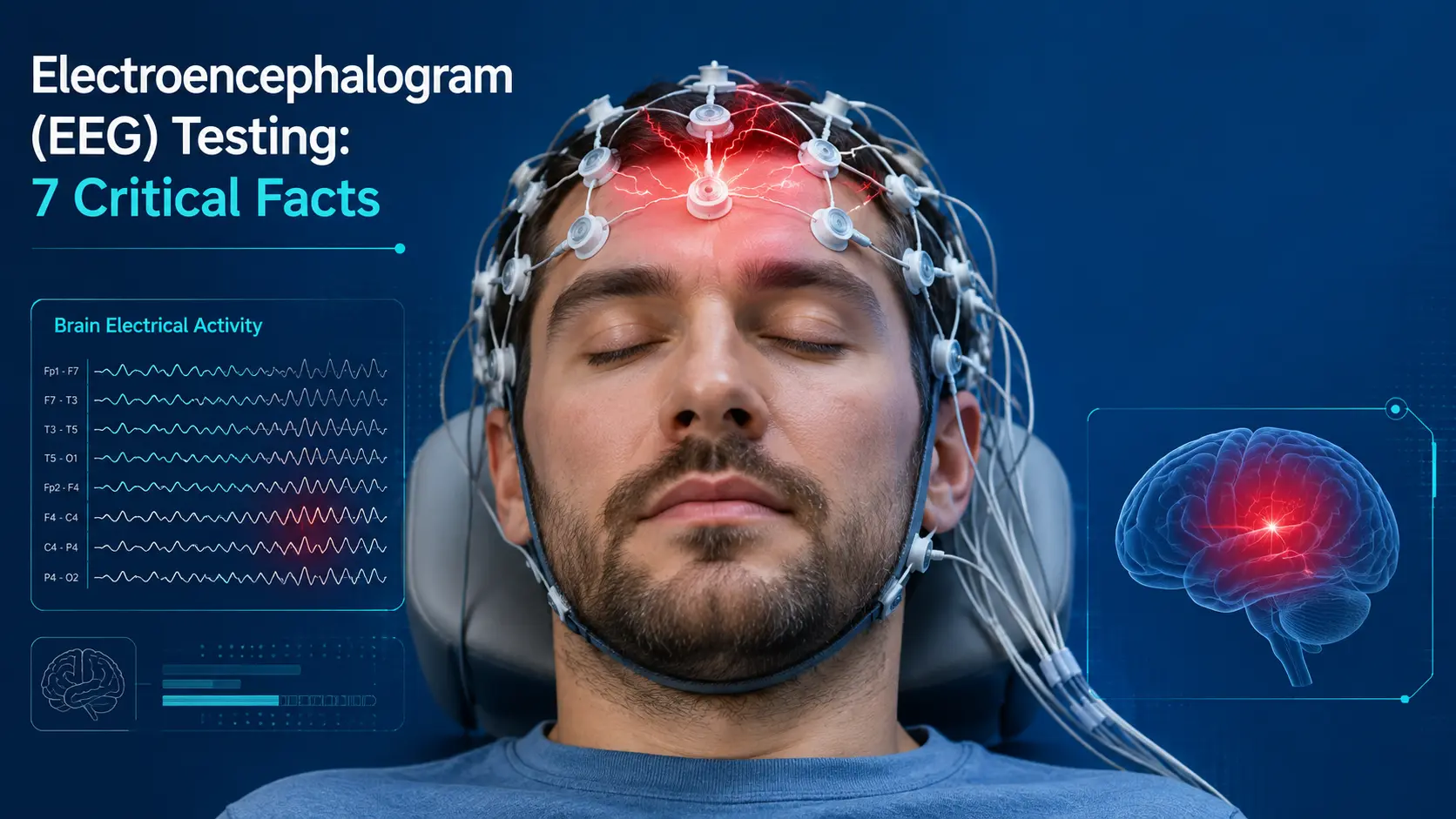
An electroencephalogram is a recording of brain activity. Neurons in the brain communicate with each other through tiny electrical impulses, and when large groups of neurons fire together, they create patterns that can be measured from the surface of the scalp. During this test, a technician uses a light adhesive paste to attach rows of small, flat metal discs called electrodes to different parts of the head. These electrodes pick up the brain’s normal electrical signals and send them to a machine, which amplifies and records them as wavy lines, often called brain waves.
Unlike an MRI or CT scan, which show the structure of the brain, this test shows brain activity in real time. It is one of the few tools doctors have that can monitor how the brain is behaving from one moment to the next, which is especially valuable for detecting abnormal electrical activity like seizures as they occur.
The procedure is completely painless. The sensors are only on the surface of the skin — nothing is inserted into the body and no electricity is sent to the brain. Most patients describe the experience as uneventful, except for the slightly unusual sensation of wearing a hat full of wires for a moment.
Why would a doctor order an electroencephalogram?
Neurologists order an electroencephalogram for a variety of reasons. This test is primarily used to diagnose epilepsy and seizure disorders, but its usefulness is not limited to these conditions.
Common reasons for EEG testing
- Diagnosing or classifying seizures and epilepsy
- Searching for the cause of unexplained fainting or confusion
- Evaluating sleep problems, including unusual nighttime behavior
- Evaluating brain activity after a severe head injury
- Monitoring brain function during and after a stroke
- Evaluating memory loss or cognitive decline
- Ascertaining brain activity levels in the critical care setting
- Searching for the cause of unexplained dizziness or vertigo
If you have experienced a loss of consciousness, our article on syncope and loss of consciousness explains how this type of brain wave test can help rule out a neurological cause. Patients recovering from a head injury are often referred for the same evaluation as part of a larger examination — you can read more about this in our guide to head injury symptoms, recovery, and treatment.
Since seizures don’t always occur as people expect, an electroencephalogram is often crucial in confirming the diagnosis. For more information on seizure activity and how it’s classified, see our post on Epilepsy and Seizures: 10 Important Facts. Those with a family history of memory changes may also benefit from our brief overview of the early signs of Alzheimer’s disease, as cognitive decline is also a common reason for this test.
How does an electroencephalogram work?
The procedure for this test is simpler than most patients expect. Here’s what typically happens during an appointment.
Step 1: Preparation
A technician will measure your head and mark the locations for the electrodes using a specific procedure, ensuring that the same areas of each patient’s brain are being measured consistently. A small amount of gel or paste is applied to each location to improve the electrical connection between the scalp and the sensors.
Step 2: Electrode placement
Usually 16 to 25 electrodes are attached to the scalp using paste, or sometimes a tight-fitting cap with sensors pre-attached. Each electrode is connected to a recording machine by a thin wire, so the setup is not as complicated as it may seem.
Step 3: Recording
Once everything is connected, you will be asked to sit or lie down comfortably and relax. The technician will record your brain’s electrical activity for a set period of time, usually between 20 and 60 minutes. You may be asked to do the following during this time:
- Close your eyes and relax
- Breathe rapidly for a few minutes (hyperventilation)
- Look at a flashing light (photic stimulation)
- Fall asleep for a while, especially for sleep-related studies
These activities are used because some abnormal electrical patterns only occur under certain conditions, so the technician needs to gently stimulate different states of the brain during the recording.
Step 4: Removal and Review
Once the recording is complete, the electrodes and paste are removed, and you can usually wash your hair immediately. A neurologist will then review the recorded brain waves and look for patterns that are outside the normal range for your age and level of alertness.
Who performs and interprets an electroencephalogram?
The electrodes are usually placed by a trained EEG technologist, sometimes called an electroneurodiagnostic technologist, who has special certification in scalp measurements and equipment operation. Once the recording is complete, a neurologist — who often has additional training in epilepsy or clinical neurophysiology — reads the tracing and prepares a written report. This combination of a skilled technologist and an experienced physician makes the results of this test reliable enough to make important medical decisions.

Types of Electroencephalogram Tests
Not all recordings are done the same way. Depending on what your doctor is trying to assess, you may be scheduled for one of several types of this test. The table below provides a brief description of the most common options.
| Type of EEG | Typical Duration | Most Commonly Used |
|---|---|---|
| Routine EEG | 20–40 minutes | General screening for seizures or unusual symptoms |
| Sleep-deprived EEG | 1–2 hours | Detects seizures that occur only with fatigue |
| Ambulatory EEG | 24–72 hours (worn at home) | Detects irregular or unexpected seizures |
| Video EEG Monitoring | 1–7 days (hospitalized) | Determines the relationship between brain wave changes and physical activity |
| Sleep EEG | Overnight | To diagnose sleep disorders and abnormal nocturnal activity |
Patients who are restless at night or have unusual movements at night are sometimes referred for a sleep-focused recording. If this sounds familiar, our article on sleep disorders and restless legs syndrome will provide helpful background before your appointment.
Ambulatory and video monitoring explained
Ambulatory recording uses a small, portable recorder that you wear at home for several days. This allows your neurologist to record brain activity during normal daily life, rather than a brief office visit. Video monitoring, on the other hand, is usually done in the hospital, where the patient’s physical movements are recorded as well as electrical observations. By monitoring the two together, doctors can determine whether a physical event, such as falling or staring, is actually related to abnormal brain activity, or whether there is another, non-neurological cause.
What to expect before, during, and after your appointment
Before the test
You will usually be asked to wash your hair the night before without using conditioner, oil, or styling products, as these can interfere with the connection to the electrodes. If you are planning to have a sleep-deprived study, your doctor may ask you to get less sleep the night before, or to continue taking your regular medications until otherwise instructed.
During the test
The recording process is quiet and passive. Most patients simply lie down and relax and follow brief instructions given by the technician. The procedure is painless, and the sensors do not send any electrical currents through the body — they simply detect signals that are generated naturally by your brain.
After the test
There are no restrictions after a typical appointment. You can drive, return to work, and resume your normal activities immediately. If you were given a sedative for a pediatric or video study, you may need someone to drive you home afterward.
Preparing Children for an EEG
Children often need a little extra reassurance before this type of test. Bringing a favorite blanket or toy, explaining the procedure in simple, non-threatening language, and letting the child watch their caregiver or sibling do a mock version of the setup — all of these can help reduce anxiety. Some pediatric centers allow parents to stay in the room during the entire recording, which makes the experience much smoother for younger patients, who might otherwise feel anxious about wires and tape.
Understanding Your Electroencephalogram (EEG) Test Results
After your neurologist reviews the recording, the results are usually described as normal or abnormal. A normal reading shows a well-organized, rhythmic pattern of brain waves that is appropriate for your age and level of alertness. An abnormal result may show spikes, sharp waves, or slowing of the waves in a specific area, which may indicate seizures, structural changes in the brain, or changes in consciousness.
It is important to note that even a single recording can sometimes seem normal in someone with epilepsy, because seizures do not occur every moment of every day. If the first test does not provide a clear answer, your doctor may recommend an extended or ambulatory study, which is one reason why.
Brain Wave Patterns Explained
EEG readings are typically divided into five wave classes based on frequency, each of which is associated with a different mental or physical condition.
| Brain Wave | Frequency Range | Associated States |
|---|---|---|
| Delta | 0.5–4 Hz | Deep, dreamless sleep |
| Theta | 4–8 Hz | Light sleep, deep relaxation |
| Alpha | 8–13 Hz | Quiet wakefulness, eyes closed |
| Beta | 13–30 Hz | Active thinking, alertness |
| Gamma | 30+ Hz | High-level information processing |
Understanding these patterns helps explain why an electroencephalogram is so useful for assessing everything from seizure disorders to sleep quality — each wave pattern tells a different part of the story about brain function.
According to the Cleveland Clinic, this type of test is safe, painless, and can be performed while the patient is awake or asleep, making it one of the most versatile tools available in neurology. Additional technical background on how these signals are generated and interpreted is available through the National Institutes of Health’s StatPearls resource on electroencephalogram testing.

Electroencephalogram vs. MRI and CT Scan
Patients often ask why a neurologist would order this test instead of or in addition to an MRI or CT scan. The short answer is that each device measures different things. An MRI or CT scan produces detailed images of the brain’s structure — revealing tumors, bleeding, or physical damage. On the other hand, such electrical recordings capture moment-by-moment brain activity, revealing how the brain’s electrical system is functioning, even when the structure appears completely normal on the scan.
Many patients ultimately need both tests: imaging to rule out structural problems and electrical monitoring to assess function. The two tests together give a neurologist a much more complete picture than either test alone. This is especially true after a head injury or stroke, when doctors want to understand both what the brain looks like and how it is currently functioning before deciding on the next steps for treatment or rehabilitation.
Is an electroencephalogram safe?
Yes. This test is considered one of the safest procedures in neurology. It is noninvasive, does not involve radiation, and has virtually no risk of complications. The only parts of the test that cause mild, temporary discomfort are the hyperventilation or flashing-lights portion, which may occasionally cause headaches or brief dizziness in sensitive individuals. Your care team will monitor you closely throughout the entire procedure and may stop these parts at any time if necessary.
How much does an electroencephalogram cost?
The cost of this type of test varies depending on your location, the type of test, and your insurance coverage. A routine outpatient test is usually much less expensive than a longer ambulatory or inpatient video recording, because the longer test requires longer use of equipment and additional doctor reviews. Many insurance plans, including Medicare and Medicaid, cover the cost of this test when a doctor deems it medically necessary. Before your appointment, our billing team can help you review your benefits to ensure there are no unexpected costs, and self-pay options are also available for those without insurance coverage.
Living with Your Results
Receiving a diagnosis after an abnormal recording can be daunting, but it’s also the first step toward getting the right treatment plan. Depending on the test results, your neurologist may recommend medication, lifestyle changes, further imaging, or a referral to a specialist. Many people with epilepsy can live full and active lives once their seizures are well controlled, and regular follow-up tests can help monitor how effective a treatment plan is over time. If your results point to a related condition, you can explore our other neurology resources — like our guide to the 24 neurological conditions we treat — to help you figure out what the next steps are.
Electroencephalogram Testing at Consultant Corner
Consultant Corner provides comprehensive electroencephalogram testing and neurological evaluations for patients across two states. Our facility is led by Saqib A. Chaudhry, MD, a board-certified vascular neurologist and neuro-intensivist, who has extensive experience interpreting complex EEG studies, managing stroke care, and treating a variety of neurological disorders.
We proudly serve patients through our two branches:
- Consultant Corner — Neurological Care in Bloomington, IL, your local neurologist in Bloomington, Illinois
- Consultant Corner — Neurological Care in El Paso, TX, your trusted health advisor in El Paso, Texas
If you would like to learn more about our doctors’ backgrounds and areas of expertise, visit our Our Doctors page, or see a full list of the conditions we treat on our Our Services page. Patients who are experiencing stroke symptoms may find our article Ischemic Stroke: Early Warning Signs, Treatment and Recovery and our Guide to TIA (Mini-Stroke) Warning Signs helpful. If you’re here because of dizziness or imbalance, our post on Vertigo and Dizziness: 9 Common Causes is another helpful resource, and our guide on 15 Signs to See a Neurology Consultant can help you schedule your first appointment.
Our Partnership with MyCare Express Care
Consultant Corner is proud to partner with MyCare Express Care, a walk-in clinic located in Bloomington, Illinois. This partnership makes it easier for our patients to access same-day evaluations, virtual neurology appointments, and integrated follow-up care between visits. MyCare Express Care is open seven days a week with extended hours and now also offers virtual neurology consultations, helping patients connect with the right specialist quickly without long waits or complicated schedules. If you need urgent, but non-urgent care in addition to your ongoing neurological treatment, their team serves as a trusted complement to the care you receive at Consultant Corner.
Frequently Asked Questions About Electroencephalogram Tests
How long does an electroencephalogram take?
A typical appointment usually takes 20 to 60 minutes, including setup and electrode removal. Extended tests, such as ambulatory or video monitoring, can last from 24 hours to a full week, depending on what your neurologist wants to observe.
Is an electroencephalogram painful?
No. This test is completely painless. The electrodes are only on the surface of the scalp and do not penetrate the skin or send any electrical current to the brain.
Can I eat or take medication before an electroencephalogram?
In most cases, yes. Unless your doctor specifically instructs you otherwise, you can eat normally and continue taking your regular medications. However, you should avoid caffeine on the day of the test, as it can affect brain wave patterns and make the results difficult to interpret.
What should I do with my hair before an EEG?
Wash your hair the night before or the morning of your appointment, but do not use conditioner, oils, gels, or sprays. Clean, product-free hair helps the electrodes connect better to your scalp.
Does an abnormal EEG result always mean that I have epilepsy?
This is not necessarily the case. An abnormal result can reflect a variety of medical conditions, including a past head injury, lack of sleep, certain medications, or metabolic changes. Your neurologist will compare the results with your symptoms and medical history before making a diagnosis.
How soon will I get my EEG results?
The time frame varies by clinic, but most patients receive a summary from their neurologist within a few days to about a week. It may take a little longer to fully review the results of complex or lengthy tests.
Can an EEG detect a stroke?
An EEG is not usually the first-line test for a suspected stroke — imaging tests like a CT or MRI are usually ordered first. However, this type of brain wave monitoring can help doctors assess ongoing brain activity later, especially in critical care settings where continuous monitoring is required.
Is an EEG covered by insurance?
Most insurance plans cover this test if it is ordered by a doctor for a medically necessary reason. Coverage details vary by plan, so it is a good idea to check with your insurance provider about your specific benefits before your appointment.
Talk to a neurologist about an EEG test
If your doctor has recommended this test, or if you are experiencing symptoms such as unexplained seizures, loss of consciousness, or memory changes, the team at Consultant Corner is ready to help you. We provide personalized, patient-centered care with advanced diagnostic testing at our Bloomington, Illinois and El Paso, Texas locations.
Ready to schedule an evaluation or have questions about electroencephalogram testing? Contact Consultant Corner online and our team will be happy to discuss your neurological health needs.