
Early Brain Health Care Can Save Lives: 12 Warning Signs
Early Brain Health Care Can Save Lives: 12 Warning Signs You Should Never Ignore Brain Health Care is not something most people think about until a symptom forces them to. Yet the truth is simple: the earlier a neurological problem is caught, the better the outcome tends to be. Many serious neurological disorders — from stroke to Alzheimer’s disease — become far more difficult to treat once diagnosis is delayed, and in some cases, that delay can mean the difference between a full recovery and permanent disability. At Consultant Corner, we see this pattern again and again. A patient waits a few days to “see if the numbness goes away.” A family assumes memory slips are “just aging.” A headache is dismissed as stress. In many cases, nothing serious is happening. But in the cases where something is wrong, that waiting period is exactly when the most damage occurs. This is why proactive brain health care — built around prompt evaluation and fast scheduling — is one of the most important things you can do for your long-term independence and quality of life. In this guide, we’ll walk through the 12 warning signs that call for immediate neurological evaluation, explain why timing matters so much in neurology, and show you how Consultant Corner supports patients across Illinois and Texas with rapid access to expert care. Why Brain Health Care Depends on Speed Unlike many areas of medicine, neurology is often a race against the clock. Brain tissue is extremely sensitive to interruptions in blood flow and oxygen, and nerve damage can become permanent within a matter of hours. This is why brain health care providers emphasize early evaluation over a “wait and see” approach. Consider ischemic stroke, one of the most time-sensitive emergencies in all of medicine. When a blood vessel supplying the brain becomes blocked, brain cells begin to die within minutes. Our detailed guide on Ischemic Stroke: Early Warning Signs, Treatment and Recovery explains how quickly treatment decisions need to be made and why every minute of delay can cost brain function that may never fully return. The same principle applies — just on a different timeline — to conditions like Alzheimer’s disease, chronic migraines, epilepsy, and nerve pain. The disease process may move slower, but early brain health care still changes the trajectory. Catching a problem in its earliest stage typically means: Fewer complications A wider range of treatment options Better response to medication and therapy Slower disease progression Greater independence over the long term The Cost of Delayed Diagnosis When neurological symptoms are ignored or misattributed to stress, aging, or “being tired,” patients often lose the window where treatment is most effective. A tremor dismissed for years as nervousness may turn out to be an early, very treatable form of essential tremor — something we cover in depth in Essential Tremor Treatment: 10 Expert Tips to Stop the Shaking. A “few dizzy spells” may be an inner-ear issue — or it may be something more serious, as explained in Vertigo & Dizziness: 9 Common Causes. The pattern is consistent: symptoms that seem minor in isolation can be early signals of a much larger issue. That is precisely why brain health care should never be reactive-only. It should be proactive. 12 Warning Signs That Call for Immediate Neurological Evaluation Below are the 12 symptoms our neurology team most frequently identifies as red flags. If you or a loved one experiences any of these, it’s time to prioritize brain health care and schedule an evaluation right away. 1. Sudden Weakness Sudden weakness, especially on one side of the body — a drooping face, a weak arm, or a leg that suddenly gives out — is one of the classic warning signs of stroke. It should always be treated as an emergency. 2. Difficulty Speaking Slurred speech, trouble finding words, or sudden confusion when trying to communicate can signal a stroke, TIA (mini-stroke), or another acute neurological event. Our article on TIA (Mini-Stroke) Warning Signs You Should Learn explains why these episodes are easy to miss — and why they demand urgent follow-up even if symptoms resolve on their own. 3. Vision Loss Sudden blurred vision, double vision, or partial vision loss in one or both eyes can point to a vascular event in the brain or an underlying neurological condition that requires prompt imaging and evaluation. 4. Severe Headaches A headache that is sudden, unusually intense (“the worst headache of your life”), or paired with other neurological symptoms is different from an everyday tension headache. Our guide, Understanding Headache / Migraine, breaks down which headache patterns are ordinary and which ones warrant a same-week appointment. 5. Confusion Sudden confusion, disorientation, or an inability to follow a conversation can be a sign of stroke, seizure activity, or a metabolic or neurological emergency. This symptom is frequently underestimated by families who assume it’s temporary. 6. Loss of Balance Trouble walking, a sudden loss of coordination, or a feeling that the room is spinning can stem from several different neurological causes. If it happens suddenly and doesn’t resolve quickly, it deserves the same urgency as other stroke symptoms. 7. Seizures A first-time seizure — even a brief one — should always prompt a full neurological workup. Recurring seizures are a hallmark of epilepsy, and early diagnosis dramatically improves how well the condition can be managed long term. 8. Memory Decline Occasional forgetfulness is normal. But memory decline that interferes with daily routines, familiar tasks, or conversations may be an early sign of Alzheimer’s disease. Our related resource, Understanding Alzheimer’s Disease, outlines the earliest changes families should watch for. 9. Frequent Falls Falling more often than usual, especially without an obvious cause, can point to problems with balance, muscle strength, or nerve signaling. Our post on 8 Muscle Weakness Signs That May Need ALS Screening explains how progressive weakness and falls can sometimes be connected to a more serious underlying condition. 10. Persistent Numbness Numbness or tingling that doesn’t go away
Brain Health Care in Texas: 10 Warning Signs
Brain Health Care in Texas: 10 Warning Signs You Shouldn’t Ignore Brain health care is something most Texans don’t think about until a symptom forces the issue — a sudden headache that won’t quit, a hand that won’t stop shaking, or a parent who suddenly can’t remember the way home. But it isn’t just for emergencies. It’s an ongoing part of staying well, and across Texas, millions of people need this kind of neurological attention every single year. As the state’s population grows and ages, neurological conditions are becoming one of the most common reasons people see a specialist. Headaches, stroke, memory loss, Parkinson’s disease, neuropathy, seizures, dizziness, and spinal nerve disorders affect people in every corner of the state, from major metro areas to small rural towns. So many of these conditions develop slowly and quietly, which is exactly why a proactive approach built around early detection can be the difference between a manageable diagnosis and a life-altering one. This guide walks through why this matters so much right now, the ten warning signs every Texan should know, a closer look at the most common neurological conditions behind them, practical guidance on when a symptom is an emergency versus something to schedule, and how to find the right specialist — no matter where in Texas you live. Along the way, we’ll also cover the diagnostic tools neurologists rely on and answer some of the questions patients ask most often. Why Brain Health Care Matters More Than Ever in Texas Texas is growing fast, and its population is aging right along with it. That combination — more people, living longer — means more neurological care needs across the board every single year. Neurological disease doesn’t discriminate by zip code, but it does become more common with age, and Texas now has one of the largest populations of older adults in the country. Rural counties face an added challenge: fewer specialists per capita, which often means longer travel times and longer waits before someone with a developing condition ever sees a neurologist. The numbers help explain why. Nationally, someone in the United States has a stroke roughly every 40 seconds, and more than 795,000 people experience one every year. Texas ranks third in the nation for the number of residents living with Alzheimer’s disease, according to the Alzheimer’s Association’s Facts and Figures report, with hundreds of thousands of Texans over 65 currently affected. Migraine, epilepsy, Parkinson’s disease, and peripheral neuropathy add millions more to that total when you count every Texan living with a chronic neurological condition. None of this means every headache is a stroke or every forgotten name is Alzheimer’s. But it does mean that regular checkups, early screening, and fast attention to new symptoms deserve the same priority Texans already give to heart health or cancer screening — and that brain health care belongs in that same conversation. 10 Warning Signs That Call for Brain Health Care Many neurological diseases don’t announce themselves with a single dramatic event. Instead, they build gradually, which is exactly why early diagnosis is so important. The table below breaks down ten common warning signs, what they can indicate, and how urgently they should be evaluated. Warning Sign What It May Indicate How Urgent? Sudden weakness (face, arm, or leg) Stroke, TIA (mini-stroke) Emergency — call 911 Persistent headaches Migraine, tension headache, or secondary causes See a doctor within days Memory loss Alzheimer’s disease, other dementia Schedule an evaluation Tremors Essential tremor, Parkinson’s disease Schedule an evaluation Numbness or tingling Neuropathy, stroke, spinal nerve compression Same-day to same-week Balance problems Vertigo, cerebellar or inner-ear disorders Schedule an evaluation Speech difficulty Stroke, TIA Emergency — call 911 Vision changes Migraine, stroke, optic nerve issues Same-day evaluation Chronic back or neck pain Spinal nerve disorders, disc disease Schedule an evaluation Muscle weakness ALS, neuropathy, muscular disorders Schedule an evaluation If a symptom appears suddenly — especially sudden weakness, speech trouble, or vision loss — treat it as an emergency. For everything else on this list, a proactive approach to your neurological health works best when it starts early, before a gradual issue becomes a permanent one. A Closer Look at the Conditions Behind These Symptoms Ischemic Stroke Ischemic stroke happens when a blood vessel supplying the brain becomes blocked, cutting off oxygen to brain tissue within minutes. It’s one of the most time-sensitive emergencies in medicine — every minute of delay costs brain cells. Our detailed guide on what causes ischemic stroke breaks down risk factors, treatment windows, and recovery, and our related post on 10 recovery tips to avoid post-stroke setbacks is worth reading for anyone supporting a loved one after a stroke. For milder, temporary versions of stroke symptoms, see our piece on 7 TIA (mini-stroke) warning signs — these “warning strokes” are a major opportunity for early intervention before a full stroke occurs. Headache and Migraine Headaches are one of the most common reasons people put off seeing a specialist, often assuming the pain will simply pass. But migraine is a distinct neurological condition, not just “a bad headache,” and it’s associated with triggers, patterns, and treatable mechanisms that a neurologist can help identify. According to the National Institute of Neurological Disorders and Stroke, migraine is also linked to a higher likelihood of other conditions, including sleep disorders and mood disorders. Read more in our post on understanding headaches and migraines. Alzheimer’s Disease and Memory Loss Memory loss is easy to dismiss as “just getting older,” but when it starts interfering with conversations, routines, or personal safety, it’s a signal worth taking seriously. Our full article on understanding Alzheimer’s disease covers early warning signs families should watch for and how a neurologist evaluates memory concerns. Early evaluation in this area can help families plan ahead, access treatment options sooner, and reduce the shock of a late-stage diagnosis. Head Injury Head injuries — from falls, car accidents, sports, or workplace incidents — can range from mild concussions to serious traumatic brain injury.
Neurological Care in Bloomington – Expert Brain Health 16 Services for Illinois
Neurological Care in Bloomington – Expert Brain Health Services for Illinois Residents Neurological Care in Bloomington, IL is becoming one of the most searched healthcare needs in central Illinois, and for good reason. Brain and nervous system disorders now affect more than 100 million Americans, and the numbers keep climbing every year. Nearly 800,000 people experience a stroke annually, more than 6 million Americans live with Alzheimer’s disease, and close to 40 million people suffer from migraine attacks. Behind every one of those numbers is a person who noticed a symptom — a headache that wouldn’t go away, a hand that suddenly went numb, a memory lapse that felt different from ordinary forgetfulness — and had to decide what to do next. If you or someone you love is searching for dependable neurological care in Bloomington, IL, this guide walks through the conditions we treat, what to expect from an evaluation, when symptoms should never be ignored, and how to connect with a board-certified vascular neurologist without unnecessary delays. Why Neurological Care in Bloomington, IL Matters for Illinois Families Central Illinois has historically had fewer neurology specialists per capita than the Chicago metro area, which means many Bloomington-area residents have had to travel long distances just to see a neurologist. That gap matters because neurological symptoms are often time-sensitive. A stroke, for example, causes the brain to lose an estimated 1.9 million neurons every minute that treatment is delayed. A seizure disorder left unmanaged can affect a person’s ability to drive or work. A tremor dismissed as “just stress” can be an early sign of a treatable movement disorder. Consultant Corner was created to close that gap by bringing accessible, evidence-based neurological care in Bloomington, IL directly to the community — with prompt scheduling, telemedicine options for patients who can’t easily travel, and a physician-led approach grounded in vascular neurology and neurocritical care experience. Conditions Treated Through Neurological Care in Bloomington, IL Below is a snapshot of the neurological conditions most commonly evaluated and managed at our Bloomington practice. This is not an exhaustive list, but it reflects the symptoms our patients bring in most often. Condition Common Warning Signs Why Timely Evaluation Matters Head Injury Confusion, headache, memory gaps after impact Undetected bleeding or swelling can worsen quickly Ischemic Stroke Sudden weakness, slurred speech, facial drooping Clot-dissolving treatment is only effective within hours TIA (Mini-Stroke) Symptoms above that resolve within minutes Often a warning sign of a full stroke to come Headache / Migraine Throbbing pain, light sensitivity, nausea Chronic migraine can be managed with the right treatment plan Alzheimer’s Disease Memory loss that disrupts daily routines Early diagnosis allows more treatment and planning options Parkinson’s Disease Tremor, slowed movement, smaller handwriting Early management can slow functional decline Multiple Sclerosis Numbness, vision changes, fatigue Delayed diagnosis can allow more nerve damage to accumulate Epilepsy / Seizures Unexplained staring spells, convulsions Untreated seizures raise injury and safety risks Neuropathy & Tremors Tingling, burning, shaking hands Often tied to an underlying, treatable cause This table only scratches the surface. Our physicians also evaluate intracerebral hemorrhage, muscle weakness, vertigo, sleep disorders, ALS-related symptoms, back pain, sciatica, carpal tunnel syndrome, cervical radiculopathy, facial pain, fainting (syncope), balance disorders, and post-stroke rehabilitation needs. Meet the Physician Behind Neurological Care in Bloomington, IL Saqib A. Chaudhry, MD, is a vascular neurologist and neuro-intensivist who leads clinical care at Consultant Corner. Vascular neurology is a subspecialty focused specifically on conditions affecting blood flow to the brain — including ischemic stroke, TIA, and intracerebral hemorrhage — while his neuro-intensivist training means he is equally prepared to manage complex, acute neurological emergencies. This combination is part of what sets our approach to neurological care in Bloomington, IL apart: patients are evaluated by a physician who understands both routine outpatient neurology and high-acuity brain emergencies. You can learn more about our full physician team on the Our Doctor page, and review our full scope of services on the Our Services page. Local Access Points for Neurological Care in Bloomington, IL Consultant Corner maintains a dedicated local presence so that Bloomington-area residents can find and reach our office easily online. You can view our verified location listing here: Neurologist in Bloomington, Illinois. Consultant Corner also operates a second location for patients further south. If you or a family member lives in or travels through Texas, you can find that office listed as Health consultant in El Paso, Texas, which provides the same standard of neurological evaluation under the Consultant Corner name. Cities and Communities Served Alongside Bloomington While our core focus is neurological care in Bloomington, IL, we regularly see patients from across central and northern Illinois, including Chicago, Springfield, Peoria, Rockford, Naperville, Aurora, Joliet, Champaign, Urbana, Decatur, Quincy, Galena, Galesburg, Alton, Belleville, Carbondale, and East St. Louis. Many of these patients choose telemedicine consultations for follow-up visits, medication management, and initial screenings, reserving in-person visits for physical exams, testing, or urgent concerns. When Symptoms Mean You Shouldn’t Wait One of the hardest parts of a neurological symptom is deciding whether it’s an emergency, a “wait and see,” or something in between. As a general rule, the following symptoms should prompt an urgent evaluation rather than a wait-and-see approach: Sudden weakness or numbness on one side of the body Slurred speech or trouble finding words A “worst headache of my life” that comes on suddenly Loss of balance, vision changes, or facial drooping A seizure that has never happened before Confusion or memory loss that appears suddenly If any of these describe what you’re experiencing right now, call 911 or go to the nearest emergency room immediately — these can be signs of a stroke or another acute neurological event, and minutes matter. According to the CDC, someone in the United States has a stroke roughly every 40 seconds, which is why stroke awareness and fast action are such a core part of our mission at Consultant Corner. The National Institute of Neurological Disorders and Stroke also notes that
Millions of Americans Live with Chronic Neurological Disorders
Millions of Americans Live with Chronic Neurological Disorders Chronic neurological disorders are far more common than most people realize. More than 100 million Americans are affected by neurological conditions ranging from migraines and neuropathy to Parkinson’s disease, multiple sclerosis, epilepsy, tremors, and chronic nerve pain. Many people ignore symptoms for years before receiving an accurate diagnosis, and that delay can mean months or even years of unnecessary suffering, missed work, and strained relationships. At Consultant Corner, we see this pattern every week. A patient mentions, almost in passing, that they’ve had “bad headaches since college” or that their hands have “always been a little shaky.” What sounds minor is often the early signature of a chronic neurological disorder that has simply never been evaluated. The goal of this article is to help you recognize the signs, understand why chronic neurological disorders are so widespread, and know exactly what to do next. Why Chronic Neurological Disorders Are So Often Missed Neurological symptoms are sneaky. They tend to build slowly, fluctuate day to day, and overlap with everyday complaints like stress, aging, or poor sleep. A tremor might be blamed on too much coffee. A memory lapse might be brushed off as “just getting older.” Chronic nerve pain might be treated only at the surface, with medication that masks the symptom instead of addressing the underlying nerve damage. The Centers for Disease Control and Prevention’s National Neurological Conditions Surveillance System (NNCSS) was created specifically because neurological disorders and conditions have such substantial, sometimes devastating, effects on millions of Americans of all ages that federal public health tracking became necessary. A more recent national analysis published in JAMA Neurology, summarized by the American Academy of Neurology, found that disorders of the nervous system impacted more than 180 million of the nearly 333 million Americans in 2021 — making neurological disease the single leading cause of health loss in the country, ahead of cardiovascular disease and cancer combined. Whatever figure you look at, the takeaway is the same: chronic neurological disorders are not rare, and they are not something to “wait out.” What Qualifies as a Chronic Neurological Disorder? A chronic neurological disorder is any condition affecting the brain, spinal cord, or peripheral nerves that persists over months or years rather than resolving on its own. Unlike an acute event — a single seizure, a one-time head injury, or a short-lived pinched nerve — chronic neurological disorders tend to be ongoing, sometimes progressive, and usually require a long-term management plan rather than a one-time fix. Here is a quick snapshot of how common some of the most frequently diagnosed chronic neurological disorders are among American adults: Condition Estimated Americans Affected Typical Age of Onset Migraine ~58 million Teens to 40s Tension-type headache ~122 million Any age Diabetic neuropathy ~17 million 40s and older Epilepsy ~3.4 million Childhood or after age 60 Parkinson’s disease ~1 million 60s and older Multiple sclerosis ~1 million 20s–40s Essential tremor Several million 40s and older Restless legs syndrome 7–10% of U.S. adults Middle age Figures reflect national epidemiological estimates and can vary by data source and year. What Increases Your Risk of a Chronic Neurological Disorder? No single factor guarantees whether someone will develop a chronic neurological disorder, but certain patterns show up again and again in patients we evaluate. Understanding these risk factors doesn’t just help with prevention — it also helps you recognize when a new symptom deserves closer attention rather than being dismissed. Age. Many chronic neurological disorders, including Parkinson’s disease and essential tremor, become more common after age 60, while others like multiple sclerosis and migraine typically appear much earlier, often between the teenage years and the early 40s. Family history. A parent or sibling with migraine, epilepsy, or essential tremor meaningfully raises the odds that a person will eventually develop a similar chronic neurological disorder themselves. Chronic health conditions. Diabetes, high blood pressure, and autoimmune disease are all closely linked to neurological complications, particularly diabetic neuropathy and stroke-related damage to the nervous system. Repeated head trauma. Concussions and head injuries, even ones that seemed minor at the time, can contribute to long-term neurological symptoms including chronic headache, memory changes, and mood disturbances. Lifestyle factors. Poor sleep, chronic stress, sedentary habits, smoking, and heavy alcohol use all appear to increase the risk or worsen the course of several chronic neurological disorders. Environmental and occupational exposures. Certain toxins, repetitive physical strain, and some workplace exposures have been associated with a higher incidence of neurological disease in long-term studies. None of these risk factors mean a chronic neurological disorder is inevitable, and none of them should be used to self-diagnose. They simply help explain why two people with the same symptom can have very different underlying causes, and why a proper neurological workup matters so much. The Most Common Chronic Neurological Disorders We Treat Consultant Corner provides evaluation and treatment for a full range of chronic neurological disorders. Below is a closer look at each condition, along with links to our in-depth guides on each topic. Headache & Migraine Chronic headache and migraine disorders are among the most underdiagnosed chronic neurological disorders in the country, largely because so many people simply reach for over-the-counter medication instead of seeking a neurological evaluation. Migraine involves much more than head pain — it can include nausea, visual disturbances, light and sound sensitivity, and even temporary weakness in severe cases. If your headaches are frequent, disabling, or changing in pattern, it’s worth a real evaluation rather than another bottle of pain relievers. Read more in our guide on Migraines: Common Triggers, Symptoms and Prevention Tips and our overview of Understanding Headache / Migraine. Treatment for this chronic neurological disorder usually starts with identifying triggers — skipped meals, dehydration, certain foods, hormonal shifts, and disrupted sleep are common culprits — and pairing that awareness with the right preventive or abortive medication. For patients with frequent or disabling migraine, newer preventive therapies have made a meaningful difference in reducing both the frequency
10 Early Alzheimer’s Disease Symptoms You Should Never Ignore
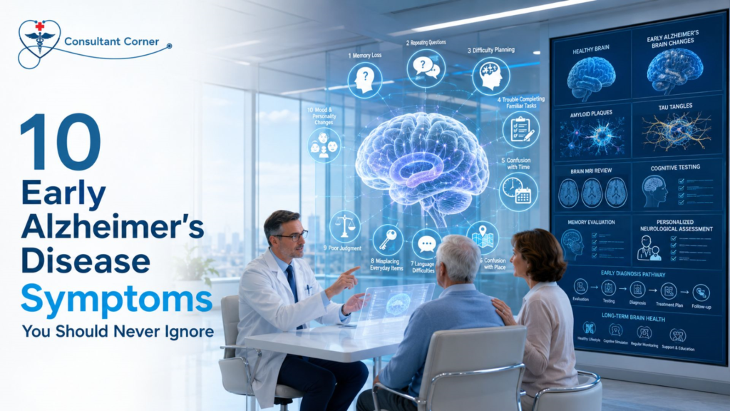
10 Early Alzheimer’s Disease Symptoms You Should Never Ignore Alzheimer’s Disease rarely announces itself all at once. It usually begins as something small — a missed appointment, a repeated question, a familiar word that suddenly feels out of reach. Because these moments look so much like ordinary forgetfulness, families often wait months or even years before asking a doctor whether something more serious might be going on. That delay matters. Recognizing Alzheimer’s Disease in its earliest stage gives patients and families more time to plan, more treatment options to consider, and a much better chance of protecting independence for as long as possible. At Consultant Corner, our neurology team evaluates memory concerns every week, and one pattern shows up again and again: the people who come in early almost always have more options than the people who wait. Below are the 10 early Alzheimer’s Disease symptoms our neurologists want every patient and caregiver to know, along with what to do if you recognize them in yourself or someone you love. It’s worth repeating that Alzheimer’s Disease is not a single dramatic event. It’s a gradual disease process, and the symptoms below can appear subtly at first — noticed only by a spouse, an adult child, or a close friend who sees the person regularly. That’s exactly why this list exists: not to cause alarm over every forgotten name, but to help you recognize the difference between ordinary aging and a pattern that deserves a professional evaluation. Why Early Symptoms of Alzheimer’s Disease Are So Easy to Miss Alzheimer’s Disease develops gradually, and the brain changes associated with it — the buildup of amyloid plaques and tau tangles — can begin a decade or more before obvious symptoms appear. In those early years, memory lapses are often chalked up to stress, poor sleep, or “just getting older.” The problem is that Alzheimer’s Disease symptoms don’t stay the same size. What starts as an occasionally misplaced set of keys can, over time, progress into disorientation in familiar places or trouble managing medications and finances. The goal isn’t to panic over every forgotten name. It’s to notice a pattern — memory or thinking changes that are frequent, progressive, and start to interfere with daily life. That pattern is the signal worth paying attention to. 1. Memory Loss That Disrupts Daily Life This is the hallmark early symptom of Alzheimer’s Disease. It looks like forgetting recently learned information, missing important dates, asking the same question multiple times in a single conversation, or relying heavily on notes and reminders for things that used to be automatic. Occasionally forgetting a name and remembering it later is normal aging. Forgetting the conversation ever happened at all is different. 2. Difficulty Planning or Solving Problems People in the early stages of Alzheimer’s Disease often struggle to follow a recipe, manage monthly bills, or keep track of a familiar process at work. Tasks that involve numbers, sequencing, or concentration take noticeably longer and require more effort than they used to. 3. Trouble Completing Familiar Tasks Driving to a well-known location, using household appliances, or remembering the rules of a favorite game can suddenly become confusing. This is different from occasionally needing help figuring out a new gadget — it’s difficulty with tasks the person has done comfortably for years. 4. Confusion With Time or Place Losing track of dates, seasons, or the passage of time is a common early Alzheimer’s Disease symptom. Someone may forget where they are, how they got there, or struggle to understand something unless it’s happening in the present moment. 5. Vision and Spatial Problems Some people with Alzheimer’s Disease develop difficulty reading, judging distances, or determining color and contrast — which can also affect driving safety. These changes are sometimes mistaken for ordinary vision decline, so an evaluation should rule out both eye conditions and neurological causes. 6. New Problems With Words in Speaking or Writing Struggling to find the right word, stopping mid-sentence, repeating phrases, or calling familiar objects by the wrong name are all early warning signs. This goes beyond the occasional “it’s on the tip of my tongue” moment everyone experiences. 7. Misplacing Items and Losing the Ability to Retrace Steps Putting things in unusual places — like keys in the refrigerator — and being unable to retrace steps to find them is a recognized early symptom of Alzheimer’s Disease. It’s often accompanied by suspicion that others have taken or moved the item. 8. Decreased or Poor Judgment Alzheimer’s Disease can affect a person’s ability to make sound decisions, whether that means falling for scams, giving away large sums of money, or paying less attention to grooming and personal hygiene than usual. 9. Withdrawal From Work or Social Activities People experiencing early memory or cognitive changes may pull back from hobbies, social events, or work projects because they’re having trouble keeping up — and because they feel self-conscious about it. This withdrawal is often one of the first things family members notice. 10. Changes in Mood and Personality Confusion, suspicion, depression, fear, and anxiety are common as Alzheimer’s Disease progresses. A person may become easily upset at home, with friends, or when taken out of a comfortable, familiar routine. Risk Factors That Increase the Chance of Alzheimer’s Disease Not everyone develops Alzheimer’s Disease, and researchers are still working to fully understand why some people are affected and others are not. That said, several risk factors are well established: Age — the single biggest known risk factor; risk rises significantly after age 65. Family history and genetics — having a parent or sibling with Alzheimer’s Disease raises personal risk, though it doesn’t guarantee a diagnosis. Cardiovascular health — high blood pressure, high cholesterol, diabetes, and obesity are all linked to higher dementia risk, likely because they affect blood flow to the brain. Head injury history — a prior head injury or concussion, especially repeated trauma, is associated with increased long-term risk. Sleep quality — chronic poor sleep or untreated sleep apnea
11 Migraine Triggers That Could Be Causing Your Headaches
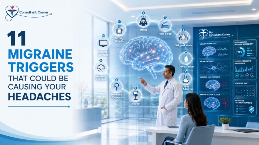
11 Migraine Triggers That Could Be Causing Your Headaches Headache & Migraine pain rarely shows up out of nowhere. For most people living with recurring attacks, an identifiable trigger — or a combination of several — sets the pain in motion hours or even a full day before the first throb hits. If you have ever wondered why one week brings three migraines and the next brings none, the answer usually isn’t bad luck. It’s a pattern hiding in your sleep schedule, your diet, your stress levels, or your environment, and once you can name it, you can start managing it. At Consultant Corner, we evaluate Headache & Migraine patients every week who are surprised to learn just how many everyday factors can set off an attack. Some triggers are obvious, like a stressful deadline or a missed meal. Others are far more subtle — a shift in barometric pressure, a whiff of perfume, or an extra hour of sleep on a Saturday morning. Because migraine triggers rarely act alone, most patients are dealing with a combination of two or three factors stacking together before an attack finally breaks through. Below, we break down the 11 most common migraine triggers, explain the science behind each one, and share practical steps you can take to reduce how often — and how severely — migraines interrupt your life. Whether you experience an occasional migraine or you’re managing a chronic pattern of frequent attacks, understanding what’s setting off your pain is the foundation of any effective treatment plan. Why Identifying Your Headache & Migraine Triggers Matters Migraine is a neurological condition, not “just a bad headache.” Research from the Mayo Clinic explains that migraine attacks can cause intense throbbing pain, nausea, and extreme sensitivity to light and sound, and they can last anywhere from a few hours to several days. Because the brain of someone with migraine disease is more sensitive to change than the average brain, small shifts — a missed meal, a poor night’s sleep, a change in the weather — can be enough to trigger a full attack. That sensitivity is exactly why trigger identification matters so much. Our team at Consultant Corner walks patients through the process of connecting the dots between lifestyle patterns and headache frequency, which is also covered in more depth on our Understanding Headache / Migraine page. A headache and migraine diary — tracking sleep, meals, stress, weather, and menstrual cycle alongside every attack — is often the single most useful tool a patient can bring to their first appointment. It’s also worth understanding that triggers and causes are not the same thing. Migraine is a genetic, neurological condition — you don’t develop it because of stress or bad habits. Triggers simply lower the threshold your brain needs to start an attack on a day when your nervous system is already primed to react. That’s why the same trigger, say a glass of red wine, might cause a migraine one week and nothing at all the next: it depends on what else is happening in your body at the time. Recognizing this interplay helps patients stop blaming themselves for “doing something wrong” and instead focus on the practical work of pattern-tracking and prevention. Below are the 11 triggers we see most often in our Headache & Migraine clinic, along with what the research says about why they matter. The 11 Most Common Migraine Triggers 1. Stress and Emotional Tension Stress is consistently ranked as the single most reported migraine trigger. When the body is under sustained pressure, it releases hormones like cortisol that can affect blood vessels and nerve signaling in the brain. Interestingly, it isn’t only stress itself that causes problems — the sudden drop in stress after a demanding week (sometimes called a “let-down migraine”) can be just as provocative. Simple stress-reduction techniques, including breathing exercises, short walks, and consistent downtime, can meaningfully reduce Headache & Migraine frequency over time. 2. Irregular Sleep Patterns Both too little sleep and too much sleep can trigger a migraine. Migraine sufferers tend to have brains that crave consistency, so going to bed and waking up at wildly different times — even on weekends — can be enough to set off an attack. According to the American Migraine Foundation, sleeping too much or too little is one of the most common triggers reported by patients. Aiming for a steady 7–8 hours each night is one of the simplest, most effective preventive habits available. 3. Skipped Meals and Dehydration Skipping meals causes blood sugar to drop, which can trigger a migraine in susceptible individuals. Dehydration works the same way — even mild fluid loss can reduce blood flow to the brain and bring on head pain. Keeping regular mealtimes and carrying water throughout the day are two of the easiest changes a Headache & Migraine patient can make. 4. Certain Foods and Food Additives Aged cheeses, processed meats containing nitrates, foods with monosodium glutamate (MSG), and artificial sweeteners are frequently reported migraine triggers. Not every migraine patient reacts to the same foods, which is why a food-and-headache journal is so valuable — it helps identify your personal pattern rather than relying on a generic “avoid this list.” 5. Caffeine and Alcohol Caffeine is a double-edged sword. A small amount can actually relieve an early migraine, but too much — or sudden caffeine withdrawal on a weekend when your routine changes — can trigger one. Alcohol, especially red wine and other beverages containing sulfites and histamines, is another well-documented trigger. Moderation and consistency (rather than complete avoidance) tend to work best for most patients. 6. Hormonal Changes Migraine affects women roughly three times more often than men, largely because of fluctuations in estrogen. Many women notice a strong connection between their menstrual cycle and Headache & Migraine attacks, with pain often striking in the days just before or during menstruation. Pregnancy, perimenopause, and hormonal medications like oral contraceptives can also shift migraine frequency and severity. 7. Weather and
12 Early Ischemic Stroke Symptoms That Could Save Your Life

12 Early Ischemic Stroke Symptoms That Could Save Your Life These are the warning signs of an ischemic stroke that deserve immediate attention. Some appear together, and some show up in isolation, but any single one of them is enough reason to call 911. 1. Sudden Numbness or Weakness on One Side The most classic sign of an ischemic stroke is a sudden loss of feeling or strength that affects only one side of the body — often the face, arm, or leg. This happens because each side of the brain controls the opposite side of the body, so a blockage on one side produces symptoms on the other. 2. Facial Drooping One side of the face may suddenly droop, feel numb, or fail to move normally when the person tries to smile. Facial drooping is one of the three checks used in the FAST test and is often the most visible sign to bystanders. 3. Arm Weakness or Drift Ask the person to raise both arms. If one arm drifts downward or cannot be lifted at all, this points toward a stroke affecting the motor pathways on one side of the brain. 4. Slurred or Garbled Speech Sudden difficulty forming words, slurring, or speech that sounds unusually thick or slow can indicate that an ischemic stroke is affecting the areas of the brain responsible for language and motor coordination of the mouth and tongue. 5. Trouble Understanding Others Some people having an ischemic stroke can speak but cannot understand what’s being said to them, or they respond with words that don’t make sense. This is a sign that a language-processing region of the brain has been affected. 6. Sudden Vision Loss or Blurriness Vision changes, including blurred vision, double vision, or a sudden loss of sight in one or both eyes, are common but frequently overlooked symptoms of an ischemic stroke, especially when they aren’t paired with obvious weakness. 7. Sudden, Severe Headache A headache that comes on abruptly and feels unlike any prior headache — often described as the “worst headache of my life” — should never be dismissed. While this is more classically linked to hemorrhagic stroke, it can also accompany certain ischemic events. 8. Dizziness or Loss of Balance A sudden inability to walk steadily, an unexplained loss of coordination, or a feeling that the room is spinning can be an early sign of an ischemic stroke affecting the brainstem or cerebellum. For a deeper look at causes of dizziness beyond stroke, see Vertigo & Dizziness: 9 Common Causes. 9. Sudden Confusion A sudden change in mental clarity — becoming disoriented, forgetting where you are, or struggling to follow a simple conversation — can be an easily missed symptom of an ischemic stroke, particularly in older adults. 10. Difficulty Walking Beyond dizziness, an ischemic stroke can cause a sudden stumble, a dragging leg, or an inability to coordinate the steps needed to walk normally, even without any pain. 11. Numbness or Tingling That Comes and Goes Symptoms that appear and then resolve within minutes can still represent a serious warning. This pattern is often the hallmark of a transient ischemic attack, a short-lived event that frequently precedes a full ischemic stroke. 12. Sudden Nausea or Vomiting With Other Symptoms On its own, nausea isn’t a stroke symptom, but when it appears alongside any of the signs above — especially dizziness, confusion, or weakness — it should raise immediate concern for an ischemic stroke rather than being written off as a stomach bug. Use the FAST Test to Recognize an Ischemic Stroke The FAST test remains the fastest, most reliable way to identify an ischemic stroke in the moment, and it doesn’t require any medical training to use. Letter What to Check What It Means F – Face Ask the person to smile Does one side of the face droop? A – Arms Ask the person to raise both arms Does one arm drift downward or feel weak? S – Speech Ask the person to repeat a simple sentence Is speech slurred, strange, or hard to understand? T – Time Note when symptoms started Call 911 immediately — do not wait or drive yourself Some hospitals now teach an expanded version, BE-FAST, which adds Balance and Eyes to catch strokes affecting coordination or vision. According to the American Stroke Association, learning and sharing the B.E. F.A.S.T. warning signs of stroke could help save a life. Either version works — what matters is acting the moment any of these checks come back abnormal. Why Fast Action Against an Ischemic Stroke Saves Lives Treatment for an ischemic stroke is highly time-dependent. When someone arrives at the hospital quickly enough, physicians may be able to use a clot-dissolving medication or a mechanical procedure to physically remove the blockage, both of which are far more effective the sooner they’re administered. The CDC notes that patients with ischemic strokes who receive clot-busting medication are more likely to recover fully or have less disability than patients who do not receive the drug, and are also less likely to need long-term care in a nursing home. This is also why a transient ischemic attack, or TIA, should never be brushed off just because the symptoms resolved on their own. A TIA is essentially a preview of a future ischemic stroke, and ignoring it removes the opportunity to intervene before a larger, more damaging event occurs. If you’ve experienced symptoms that came and went, it’s worth reading our full breakdown of TIA warning signs to understand how closely the two conditions are connected. Ischemic Stroke vs. Hemorrhagic Stroke Not all strokes are caused by a blockage. Understanding the difference helps explain why treatment approaches vary so much between the two. Feature Ischemic Stroke Hemorrhagic Stroke Cause Blocked artery (clot or plaque) Ruptured blood vessel Frequency About 87% of all strokes About 13% of all strokes Common Treatment Clot-dissolving medication, clot removal Surgery, blood pressure control Key Risk Factors High blood pressure, high cholesterol, atrial
10 Warning Signs of a Head Injury You Should Never Ignore
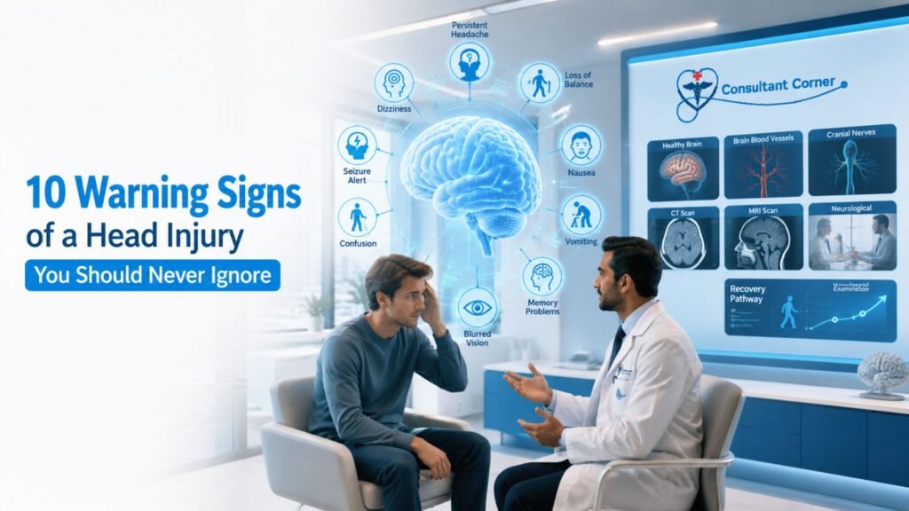
10 Warning Signs of a Head Injury You Should Never Ignore A head injury can look harmless one moment and turn serious the next. You bump your head getting out of the car, you take a fall on the stairs, or you’re in a minor fender-bender — and you feel fine. Then, hours or even days later, a headache creeps in, your thinking feels foggy, or the room starts to spin. This delayed pattern is exactly why this kind of injury is so easy to underestimate and so important to take seriously. At Consultant Corner, our neurology team evaluates patients with a head injury every week, from athletes and weekend warriors to older adults who’ve had a simple fall at home. Below, we walk through the 10 warning signs you should never ignore, explain why each one matters, and outline exactly when a bump on the head needs a same-week neurology visit — or an emergency room. Why a Head Injury Deserves Immediate Attention Every head injury is different because every brain responds to trauma differently. A blow to the head can range from a mild bump that resolves on its own to a traumatic brain injury (TBI) that causes swelling, bleeding, or lasting damage. According to the Centers for Disease Control and Prevention, some signs of a serious brain injury appear immediately, while others don’t show up until hours or days later — which is exactly why “I feel okay right now” isn’t the same as “I’m in the clear.” The danger with this kind of trauma is that the brain is a soft, delicate organ cushioned inside a hard skull. A sudden jolt, blow, or rapid deceleration can cause the brain to move within the skull, stretching nerve fibers and, in more serious cases, causing bruising or bleeding. That’s why doctors urge anyone who has experienced this kind of trauma — even a “minor” bump — to watch closely for the warning signs described below. Common Causes of a Head Injury Understanding how these injuries typically happen can help you recognize your own risk level. The most frequent causes include: Falls — the leading cause across all age groups, especially among young children and older adults Motor vehicle collisions — including car, motorcycle, and bicycle accidents Sports and recreational activities — contact sports like football, hockey, and soccer, as well as cycling and skiing Workplace accidents — falling objects, equipment accidents, or falls from height Physical altercations or assaults Regardless of the cause, the same principle applies: any forceful blow, jolt, or sudden stop that moves the brain inside the skull has the potential to cause lasting harm, so the mechanism of injury matters less than how closely you monitor your symptoms afterward. How to Reduce Your Risk of a Head Injury While not every accident can be prevented, several everyday habits meaningfully lower your risk: Wear a properly fitted helmet for biking, skiing, contact sports, and motorcycle riding Use seatbelts and appropriate child car seats on every trip, even short ones Remove fall hazards at home, such as loose rugs, poor lighting, and cluttered stairways Install handrails and grab bars for older adults or anyone with balance concerns Follow return-to-play guidelines after a prior concussion instead of rushing back into sports These small precautions won’t eliminate risk entirely, but they meaningfully reduce both the likelihood and severity of trauma to the head. Children, Older Adults, and Head Injury Risk Two groups deserve extra caution after any blow to the head: young children and older adults. Kids may not be able to describe symptoms like fogginess or light sensitivity, so parents should watch for excessive crying, unusual sleepiness, or refusal to eat or nurse. Older adults, especially those taking blood thinners, face a higher risk of bleeding inside the skull even after a seemingly minor fall, and symptoms can be subtler — mistaken for normal fatigue or confusion rather than a medical emergency. If a child or an older family member has taken a fall or a hit to the head, it’s worth erring on the side of caution and scheduling an evaluation, even if they seem fine at first. 10 Warning Signs of a Head Injury You Should Never Ignore 1. A Headache That Won’t Go Away or Keeps Getting Worse A dull ache after a bump is common, but a headache that intensifies over hours, rather than fading, is one of the most important warning signs on this list. A worsening headache can point to swelling or bleeding building up pressure inside the skull. This is different from a typical tension headache, which usually stays mild and steady rather than escalating. 2. Dizziness, Loss of Balance, or Feeling “Off” Struggling to walk a straight line, feeling unsteady on your feet, or sensing that something is generally “off” after trauma to the head often signals that the impact has disrupted the brain pathways responsible for coordination and spatial awareness. This can appear right away or develop over the following day. 3. Nausea or Repeated Vomiting Occasional queasiness after a jolt to the head isn’t unusual, but nausea that persists — or vomiting more than once — is a recognized sign of increased pressure inside the brain and should never be brushed off, especially in children and older adults. 4. Trouble Concentrating, Remembering, or Processing Information Many people describe this as feeling “foggy,” mentally slow, or unable to hold a thought after a blow to the head. Difficulty concentrating at work, forgetting recent conversations, or struggling to follow a simple task are classic post-concussion symptoms that deserve a proper neurological evaluation. 5. Sensitivity to Light or Blurry Vision Photophobia (light sensitivity) and visual disturbances such as blurred or double vision can appear immediately or emerge days after the trauma. These symptoms suggest the brain is still irritated from the impact and hasn’t finished healing. 6. Sudden, Severe Headache Unlike Any Before A “thunderclap” headache — one that comes on suddenly and feels far more intense than any
10 Recovery Tips to Avoid Post-Stroke Setbacks
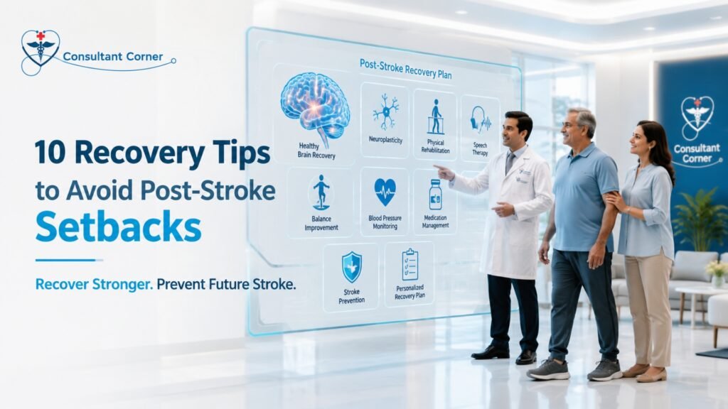
10 Recovery Tips to Avoid Post-Stroke Setbacks Post-Stroke recovery doesn’t end when you leave the hospital — it’s a daily process, and small mistakes in those first weeks and months can quietly undo the progress you’ve worked hard to make. Whether you or someone you love is a few days or several months into healing, understanding how to protect that progress is one of the most important things you can do. Below, we walk through ten practical, medically informed tips that can help you avoid the most common post-stroke setbacks and keep recovery moving in the right direction. A stroke is a sudden, life-altering event, but what happens after the hospital discharge often determines the long-term outcome. Weakness, coordination issues, balance problems, memory changes, fatigue, speech difficulty, and emotional swings can all persist for weeks, months, or years. The encouraging news is that with the right follow-up care, structured rehabilitation, and a few key lifestyle adjustments, most patients recover faster, stronger, and safer than they expect. At Consultant Corner, our neurology team has guided hundreds of patients through this critical recovery window, and we’ve seen firsthand which habits accelerate healing — and which ones quietly cause setbacks. This guide brings those lessons together in one place. No two recoveries look exactly alike, and comparing your own timeline to someone else’s story online can do more harm than good. Some patients regain independence within a few months, while others need a year or more of structured therapy before they feel like themselves again. What almost every successful recovery has in common, though, is a consistent, well-coordinated care plan — one that treats the body, the brain, and the emotional side of healing as equally important. That’s the philosophy behind every tip below. Why the Post-Stroke Recovery Window Matters So Much The brain continues to heal and rewire itself for 12 to 24 months after a stroke, a process doctors call neuroplasticity. During this period, the brain is unusually responsive to therapy, repetition, and structured stimulation. Without proper guidance, however, patients often experience slow recovery, persistent weakness, a higher risk of falls, unmanaged spasticity, and — in the most serious cases — a repeat stroke. This is exactly why Post-Stroke follow-up care isn’t optional; it’s the foundation that everything else is built on. According to the Centers for Disease Control and Prevention, recovery time after a stroke differs for everyone and can take weeks, months, or even years, with some people recovering fully while others face long-term or lifelong disabilities. That variability is precisely why an individualized recovery plan — not a generic checklist — matters so much. 10 Recovery Tips to Avoid Post-Stroke Setbacks 1. Never Skip Your Neurology Follow-Up Appointments It’s tempting to feel like you’re “done” with medical care once you’re home, but Post-Stroke follow-up visits are where your care team catches subtle changes before they become major setbacks. Regular evaluation of strength, balance, speech, and cognition allows your neurologist to adjust medications, therapy intensity, and safety recommendations as your brain heals. Patients who skip these visits are far more likely to experience preventable complications, from unmanaged spasticity to missed signs of a second stroke. 2. Stick to Your Rehabilitation Schedule — Even on Hard Days Physical, occupational, and speech therapy are not optional extras; they are the engine of Post-Stroke recovery. The brain rebuilds neural pathways through repetition, so missing sessions — even a few — can slow momentum that took weeks to build. On days when fatigue or frustration makes therapy feel impossible, talk to your care team about modifying the session rather than skipping it entirely. Consistency, not intensity, is what drives long-term gains. 3. Manage Blood Pressure, Cholesterol, and Blood Sugar Closely One of the most common causes of a second stroke is poorly controlled cardiovascular risk factors. High blood pressure, elevated cholesterol, and unmanaged diabetes all place ongoing strain on the blood vessels that supply the brain. Home monitoring, consistent medication use, and regular lab work are simple habits that meaningfully lower the risk of another event during Post-Stroke recovery. It helps to think of these numbers as an early warning system rather than an inconvenience. A home blood pressure cuff, a simple daily log, and a shared spreadsheet or notebook with your care team can catch a dangerous trend weeks before it becomes an emergency. Diet also plays a bigger role than most patients expect — reducing sodium, limiting processed foods, and increasing fiber and lean protein intake can measurably improve blood pressure and cholesterol numbers within a few months. Ask your neurologist or a referred dietitian for a plan tailored to your specific risk factors, since generic advice rarely accounts for medication interactions or kidney function. 4. Address Swallowing and Speech Difficulties Early Speech and swallowing problems (dysphagia) are common after a stroke and are often underreported by patients who feel embarrassed or assume the issue will resolve on its own. Left untreated, swallowing difficulties can lead to choking, aspiration pneumonia, and malnutrition — all of which can dramatically derail recovery. A speech-language pathologist can assess swallowing safety and provide targeted exercises that improve both speech clarity and swallowing function over time. 5. Reduce Fall Risk at Home Balance and coordination problems make falls one of the most frequent post-stroke setbacks, and a single fall can result in a fracture or head injury that resets months of progress. Simple home modifications — removing loose rugs, installing grab bars in the bathroom, improving lighting, and using assistive devices as recommended — go a long way. If dizziness or vertigo has been part of your recovery, it’s worth discussing with your care team, since balance issues sometimes overlap with other neurological conditions. A physical or occupational therapist can also perform a formal fall-risk assessment, walking through your home environment room by room to flag specific hazards you might not notice day to day — a slick bathroom floor, a poorly lit hallway, or stairs without a secure handrail. Footwear matters too; loose slippers
8 Muscle Weakness Signs That May Need ALS Screening
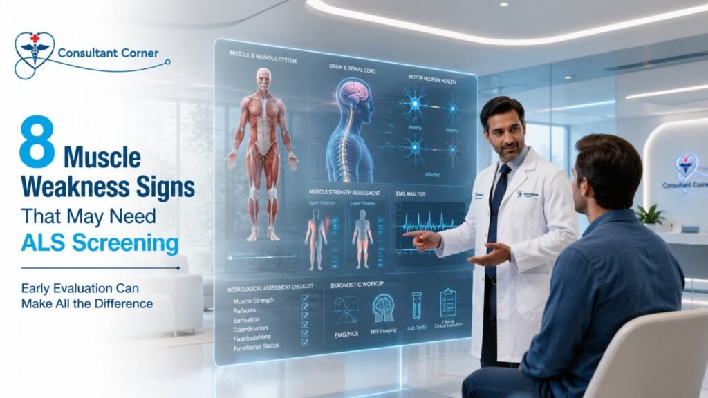
8 Muscle Weakness Signs That May Need ALS Screening Feeling weaker than usual? Losing strength in your arms, legs, or hands? Don’t ignore it — early evaluation can make all the difference. Muscle weakness is one of the most common reasons patients schedule a neurology appointment, and in the overwhelming majority of cases, it is caused by something treatable — a pinched nerve, a vitamin deficiency, thyroid dysfunction, or a compressed disc. But when muscle weakness becomes progressive, spreads from one area of the body to another, or shows up alongside twitching and unexplained fatigue, it is important to rule out rarer but more serious neurological causes, including amyotrophic lateral sclerosis (ALS). At Consultant Corner, muscle weakness and ALS screening are evaluated with the same level of urgency and precision. Our goal is simple: identify the true cause of your weakness quickly, rule out what it isn’t, and build a clear plan forward — whether that means treating a reversible condition or connecting you with the right specialists for further workup. This guide walks through eight muscle weakness signs that deserve a closer look, explains what typically causes muscle weakness, and outlines what a thorough ALS screening actually involves. Why Muscle Weakness & ALS Screening Matters Most people who notice new weakness assume the worst, and that fear alone can keep people from seeking care. The truth is reassuring: the vast majority of muscle weakness has nothing to do with ALS. Nerve compression, neuropathy, and metabolic issues account for the majority of weakness complaints seen in neurology clinics. Still, because ALS is a progressive disease where early diagnosis and early treatment meaningfully affect outcomes, muscle weakness and ALS screening should never be delayed when the warning signs listed below are present. Screening does two things at once. First, it identifies and treats the common, reversible causes of weakness that are affecting most patients. Second, it catches the small subset of cases where early neurological disease is present, so treatment can begin as soon as possible. 8 Warning Signs of Muscle Weakness That Warrant Evaluation If you’re noticing any of the following, it’s worth scheduling a muscle weakness and ALS screening evaluation rather than waiting to see if it improves on its own. 1. One Arm or Leg Becoming Progressively Weaker Weakness that is limited to one side of the body — and that continues to get worse over weeks or months rather than staying stable — is one of the more notable patterns doctors look for. Weakness that stays the same for months and doesn’t spread is far less concerning than weakness that keeps advancing. 2. Difficulty Lifting Objects or Gripping Items Trouble opening jars, holding a coffee cup, or gripping a steering wheel can point to problems ranging from carpal tunnel syndrome to cervical radiculopathy to early motor neuron changes. A hand-strength decline that shows up on both sides equally is usually different — and typically less urgent — than weakness isolated to one hand. 3. Foot Drop or Frequent Tripping Foot drop happens when the muscles that lift the front of the foot weaken, causing the toes to catch on the ground while walking. It’s a hallmark of peroneal nerve compression, but it can also be an early sign of more diffuse motor neuron involvement, which is why it’s included in a thorough muscle weakness and ALS screening. 4. Trouble Raising Your Arms or Climbing Stairs Weakness that affects the muscles closest to the body — shoulders, hips, and thighs — rather than the hands and feet has a different pattern of causes, including myopathies and certain autoimmune muscle diseases. Struggling to reach overhead or push up from a chair are both signals worth mentioning to your doctor. 5. Muscle Twitching (Fasciculations) Occasional muscle twitches, especially in the calves or eyelids, are extremely common and are almost always benign, often related to caffeine, stress, dehydration, or fatigue. Twitching that is widespread, persistent, and paired with muscle weakness is the combination that warrants a closer look during ALS screening — twitching alone, without accompanying weakness, is rarely a red flag. 6. Unexplained Fatigue With Simple Tasks Feeling unusually exhausted after routine activities — climbing a single flight of stairs, carrying groceries, or brushing your hair — can reflect anything from anemia and thyroid disease to early neuromuscular weakness. Because fatigue overlaps with so many common conditions, it’s one of the harder symptoms to interpret without proper testing. 7. Handwriting Becoming Smaller or Shakier A noticeable change in handwriting — letters shrinking, lines becoming less steady, or grip fatigue mid-sentence — can reflect fine motor weakness in the hand. While this symptom is also associated with Parkinson’s disease, in the context of progressive limb weakness it belongs on the muscle weakness and ALS screening checklist. 8. Slurred Speech, Trouble Swallowing, or Voice Changes When weakness begins to affect the muscles used for speaking or swallowing — a nasal-sounding voice, slurred words, or choking on liquids — this is one of the symptoms that should prompt an immediate evaluation, since it can also signal a stroke in its acute form. Any sudden change here deserves urgent attention. What Causes Muscle Weakness? (Most of It Isn’t ALS) It bears repeating: most muscle weakness is not ALS. A thorough muscle weakness and ALS screening looks at the full range of possible causes, including: Pinched nerves and nerve entrapment (such as carpal tunnel syndrome) Peripheral neuropathy Cervical or lumbar radiculopathy (nerve root compression in the neck or lower back) Stroke or transient ischemic attack (TIA)-related weakness Myopathies (primary muscle diseases) Thyroid disorders, including hypothyroidism Vitamin deficiencies, particularly B12 and vitamin D Autoimmune neuromuscular conditions, such as myasthenia gravis Medication side effects, including certain statins and steroids Because so many of these conditions overlap in how they present, an evaluation for muscle weakness and ALS screening is designed to systematically rule each of them in or out — not jump straight to worst-case scenarios. Who Is at Higher Risk for ALS? Understanding risk factors